2
views
0
recommends
+1 Recommend
0 collections
    0
    shares
      • Record: found
      • Abstract: found
      • Article: found
      Is Open Access

      MiR-19b-3p regulated by BC002059/ABHD10 axis promotes cell apoptosis in myocardial infarction

      research-article

      Read this article at

      Bookmark
          There is no author summary for this article yet. Authors can add summaries to their articles on ScienceOpen to make them more accessible to a non-specialist audience.

          Abstract

          Background

          Recently, microRNAs (miRNAs), have been extensively investigated in diseases. The upregulated expression of miR-19b-3p has been validated in patients with hypertrophic cardiomyopathy. Nonetheless, it regulatory mechanism in myocardial infarction (MI) is still unclear.

          Purpose

          This research aimed to investigate the role and molecular regulation mechanism of miR-19b-3p in MI.

          Methods

          QRT-PCR and western blot assays measured RNA and protein expression. Cell apoptosis were tested by flow cytometry and TUNEL assays. Cell viability was measured by trypan blue staining method. RIP and luciferase report assays examined gene interaction. The assays were performed under hypoxia condition.

          Results

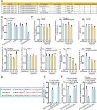
          MiR-19b-3p was highly expressed in myocardial cell line H9C2, primary cardiomyocytes, and tissues from MI mouse model. MiR-19b-3p inhibition suppressed the apoptosis of cardiomyocytes. BC002059 could up-regulate ABHD10 through sequestering miR-19b-3p. BC002059 upregulation was observed to repress cell apoptosis. Rescue experiments demonstrated that miR-19b-3p overexpression abrogated the suppressive impact of BC002059 on the apoptosis of MI cells, and infarct size, area at risk as well as CK-MB and LDH release of MI mouse model tissues, which was further abolished via ABHD10 increment.

          Conclusion

          MiR-19b-3p regulated by BC002059/ABHD10 axis promotes cell apoptosis in MI, which might provide a novel perspective for MI alleviation research.

          Related collections

          Most cited references31

          • Record: found
          • Abstract: found
          • Article: not found

          miR-133 and miR-30 regulate connective tissue growth factor: implications for a role of microRNAs in myocardial matrix remodeling.

          The myocardium of the failing heart undergoes a number of structural alterations, most notably hypertrophy of cardiac myocytes and an increase in extracellular matrix proteins, often seen as primary fibrosis. Connective tissue growth factor (CTGF) is a key molecule in the process of fibrosis and therefore seems an attractive therapeutic target. Regulation of CTGF expression at the promoter level has been studied extensively, but it is unknown how CTGF transcripts are regulated at the posttranscriptional level. Here we provide several lines of evidence to show that CTGF is importantly regulated by 2 major cardiac microRNAs (miRNAs), miR-133 and miR-30. First, the expression of both miRNAs was inversely related to the amount of CTGF in 2 rodent models of heart disease and in human pathological left ventricular hypertrophy. Second, in cultured cardiomyocytes and fibroblasts, knockdown of these miRNAs increased CTGF levels. Third, overexpression of miR-133 or miR-30c decreased CTGF levels, which was accompanied by decreased production of collagens. Fourth, we show that CTGF is a direct target of these miRNAs, because they directly interact with the 3' untranslated region of CTGF. Taken together, our results indicate that miR-133 and miR-30 importantly limit the production of CTGF. We also provide evidence that the decrease of these 2 miRNAs in pathological left ventricular hypertrophy allows CTGF levels to increase, which contributes to collagen synthesis. In conclusion, our results show that both miR-133 and miR-30 directly downregulate CTGF, a key profibrotic protein, and thereby establish an important role for these miRNAs in the control of structural changes in the extracellular matrix of the myocardium.
            Bookmark
            • Record: found
            • Abstract: found
            • Article: not found

            Long-term trends in the incidence of and survival with heart failure.

            Heart failure is a major public health problem. Long-term trends in the incidence of heart failure and survival after its onset in the community have not been characterized. We used statistical models to assess temporal trends in the incidence of heart failure and Cox proportional-hazards regression to evaluate survival after the onset of heart failure among subjects in the Framingham Heart Study. Cases of heart failure were classified according to the date of onset: 1950 through 1969 (223 cases), 1970 through 1979 (222), 1980 through 1989 (307), and 1990 through 1999 (323). We also calculated 30-day, 1-year, and 5-year age-adjusted mortality rates for each period. Heart failure occurred in 1075 subjects (51 percent of whom were women). As compared with the rate for the period from 1950 through 1969, the incidence of heart failure remained virtually unchanged among men in the three subsequent periods but declined by 31 to 40 percent among women (rate ratio for the period from 1990 through 1999, 0.69; 95 percent confidence interval, 0.51 to 0.93). The 30-day, 1-year, and 5-year age-adjusted mortality rates among men declined from 12 percent, 30 percent, and 70 percent, respectively, in the period from 1950 through 1969 to 11 percent, 28 percent, and 59 percent, respectively, in the period from 1990 through 1999. The corresponding rates among women were 18 percent, 28 percent, and 57 percent for the period from 1950 through 1969 and 10 percent, 24 percent, and 45 percent for the period from 1990 through 1999. Overall, there was an improvement in the survival rate after the onset of heart failure of 12 percent per decade (P=0.01 for men and P=0.02 for women). Over the past 50 years, the incidence of heart failure has declined among women but not among men, whereas survival after the onset of heart failure has improved in both sexes. Factors contributing to these trends need further clarification. Copyright 2002 Massachusetts Medical Society
              Bookmark
              • Record: found
              • Abstract: found
              • Article: not found

              MicroRNA-103/107 Regulate Programmed Necrosis and Myocardial Ischemia/Reperfusion Injury Through Targeting FADD.

              Necrosis is one of the main forms of cardiomyocyte death in heart disease. Recent studies have demonstrated that certain types of necrosis are regulated and programmed dependent on the activation of receptor-interacting serine/threonine-protein kinase (RIPK) 1 and 3 which may be negatively regulated by Fas-associated protein with death domain (FADD). In addition, microRNAs and long noncoding RNAs have been shown to play important roles in various biological processes recently.
                Bookmark

                Author and article information

                Contributors
                1476870009@qq.com
                78249073@qq.com
                Journal
                Biol Direct
                Biol Direct
                Biology Direct
                BioMed Central (London )
                1745-6150
                18 August 2022
                18 August 2022
                2022
                : 17
                : 20
                Affiliations
                [1 ]GRID grid.440218.b, ISNI 0000 0004 1759 7210, Department of Cardiology, , Shenzhen People’s Hospital, Second Clinical Medical College of Jinan University, ; 1017 Dongmen North Road, Luohu District, Shenzhen, 518000 Guangdong China
                [2 ]GRID grid.440218.b, ISNI 0000 0004 1759 7210, Department of Gastroenterology, , Shenzhen People’s Hospital, Second Clinical Medical College of Jinan University, ; 1017 Dongmen North Road, Luohu District, Shenzhen, 518000 Guangdong China
                Article
                333
                10.1186/s13062-022-00333-x
                9386969
                35978367
                a0c0719a-185d-410c-89bd-849a3bb1addb
                © The Author(s) 2022

                Open AccessThis article is licensed under a Creative Commons Attribution 4.0 International License, which permits use, sharing, adaptation, distribution and reproduction in any medium or format, as long as you give appropriate credit to the original author(s) and the source, provide a link to the Creative Commons licence, and indicate if changes were made. The images or other third party material in this article are included in the article's Creative Commons licence, unless indicated otherwise in a credit line to the material. If material is not included in the article's Creative Commons licence and your intended use is not permitted by statutory regulation or exceeds the permitted use, you will need to obtain permission directly from the copyright holder. To view a copy of this licence, visit http://creativecommons.org/licenses/by/4.0/. The Creative Commons Public Domain Dedication waiver ( http://creativecommons.org/publicdomain/zero/1.0/) applies to the data made available in this article, unless otherwise stated in a credit line to the data.

                History
                : 15 April 2022
                : 23 July 2022
                Funding
                Funded by: the Shenzhen Science and Technology Project
                Award ID: JCYJ20160422170251848
                Categories
                Research
                Custom metadata
                © The Author(s) 2022

                Life sciences
                myocardial infarction,apoptosis,mir-19b-3p,bc002059/abhd10 axis
                Life sciences
                myocardial infarction, apoptosis, mir-19b-3p, bc002059/abhd10 axis

                Comments

                Comment on this article