15
views
0
recommends
+1 Recommend
1 collections
    0
    shares

      International Journal of COPD (submit here)

      This international, peer-reviewed Open Access journal by Dove Medical Press focuses on pathophysiological processes underlying Chronic Obstructive Pulmonary Disease (COPD) interventions, patient focused education, and self-management protocols. Sign up for email alerts here.

      39,063 Monthly downloads/views I 2.893 Impact Factor I 5.2 CiteScore I 1.16 Source Normalized Impact per Paper (SNIP) I 0.804 Scimago Journal & Country Rank (SJR)

      • Record: found
      • Abstract: found
      • Article: found
      Is Open Access

      P-wave indices in patients with pulmonary emphysema: do P-terminal force and interatrial block have confounding effects?

      research-article

      Read this article at

      ScienceOpenPublisherPMC
      Bookmark
          There is no author summary for this article yet. Authors can add summaries to their articles on ScienceOpen to make them more accessible to a non-specialist audience.

          Abstract

          Introduction

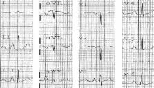
          Pulmonary emphysema causes several electrocardiogram changes, and one of the most common and well known is on the frontal P-wave axis. P-axis verticalization (P-axis > 60°) serves as a quasidiagnostic indicator of emphysema. The correlation of P-axis verticalization with the radiological severity of emphysema and severity of chronic obstructive lung function have been previously investigated and well described in the literature. However, the correlation of P-axis verticalization in emphysema with other P-indices like P-terminal force in V 1 (Ptf), amplitude of initial positive component of P-waves in V 1 (i-PV1), and interatrial block (IAB) have not been well studied. Our current study was undertaken to investigate the effects of emphysema on these P-wave indices in correlation with the verticalization of the P-vector.

          Materials and methods

          Unselected, routinely recorded electrocardiograms of 170 hospitalized emphysema patients were studied. Significant Ptf (s-Ptf) was considered ≥40 mm.ms and was divided into two types based on the morphology of P-waves in V 1: either a totally negative (−) P wave in V 1 or a biphasic (+/−) P wave in V 1.

          Results

          s-Ptf correlated better with vertical P-vectors than nonvertical P-vectors ( P = 0.03). s-Ptf also significantly correlated with IAB ( P = 0.001); however, IAB and P-vector verticalization did not appear to have any significant correlation ( P = 0.23). There was a very weak correlation between i-PV1 and frontal P-vector ( r = 0.15; P = 0.047); however, no significant correlation was found between i-PV1 and P-amplitude in lead III ( r = 0.07; P = 0.36).

          Conclusion

          We conclude that increased P-tf in emphysema may be due to downward right atrial position caused by right atrial displacement, and thus the common assumption that increased P-tf implies left atrial enlargement should be made with caution in patients with emphysema. Also, the lack of strong correlation between i-PV1 and P-amplitude in lead III or vertical P-vector may suggest the predominant role of downward right atrial distortion rather than right atrial enlargement in causing vertical P-vector in emphysema.

          Most cited references19

          • Record: found
          • Abstract: found
          • Article: not found

          Interatrial blocks. A separate entity from left atrial enlargement: a consensus report.

          Impaired interatrial conduction or interatrial block is well documented but is not described as an individual electrocardiographic (ECG) pattern in most of ECG books, although the term atrial abnormalities to encompass both concepts, left atrial enlargement (LAE) and interatrial block, has been coined. In fact, LAE and interatrial block are often associated, similarly to what happens with ventricular enlargement and ventricular block. The interatrial blocks, that is, the presence of delay of conduction between the right and left atria, are the most frequent atrial blocks. These may be of first degree (P-wave duration >120 milliseconds), third degree (longer P wave with biphasic [±] morphology in inferior leads), and second degree when these patterns appear transiently in the same ECG recording (atrial aberrancy). There are evidences that these electrocardiographic P-wave patterns are due to a block because they may (a) appear transiently, (b) be without associated atrial enlargement, and (c) may be reproduced experimentally. The presence of interatrial blocks may be seen in the absence of atrial enlargement but often are present in case of LAE. The most important clinical implications of interatrial block are the following: (a) the first degree interatrial blocks are very common, and their relation with atrial fibrillation and an increased risk for global and cardiovascular mortality has been demonstrated; (b) the third degree interatrial blocks are less frequent but are strong markers of LAE and paroxysmal supraventricular tachyarrhythmias. Their presence has been considered a true arrhythmological syndrome. Copyright © 2012 Elsevier Inc. All rights reserved.
            Bookmark
            • Record: found
            • Abstract: found
            • Article: not found

            Electromechanical dysfunction of the left atrium associated with interatrial block.

            Our purpose was to determine the effect of interatrial block (IAB, P-wave duration >/=120 ms) on left atrial (LA) dynamics. IAB is associated with LA enlargement (LAE). LA dysfunction is associated with decreased left ventricular filling, a propensity for LA appendage thrombus formation, and reduced atrial natriuretic peptide levels. We evaluated LA function in patients with and without IAB matched for LA size. Echocardiograms with LA enlargement were analyzed. Twenty-four patients had IAB, and 16 patients without IAB formed the control group. LA volumes, A-wave acceleration times (At), LA stroke volume (LASV), ejection fraction (LAEF), and kinetic energy (LAKE) were calculated. The control group and patients with IAB had comparable maximal LA volume and diameter (P >.05). Patients with IAB had significantly longer At (115 +/- 39 ms vs 83 +/- 24 ms, P <.01) and smaller LASV (7 +/- 5 mL vs 17 +/- 6 mL, P <.01), LAEF (9% +/- 6% vs 25% +/- 8%, P <.01), and LAKE (20 +/- 14 vs 65 +/- 44 Kdyne/cm/s, P <.01). LAKE varied inversely with P-wave duration (r = -0.51, P <.01). P-wave duration and minimal LA volume were independent determinants of LAEF. Patients with IAB have a sluggish, poorly contractile LA, and the extent of dysfunction is related to the degree of electrical delay from IAB. IAB should be considered a marker of an electromechanically dysfunctional LA and hence a risk factor for atrial fibrillation and congestive heart failure.
              Bookmark
              • Record: found
              • Abstract: not found
              • Article: not found

              Prevalence of interatrial block in a general hospital population.

                Bookmark

                Author and article information

                Journal
                Int J Chron Obstruct Pulmon Dis
                Int J Chron Obstruct Pulmon Dis
                International Journal of COPD
                International Journal of Chronic Obstructive Pulmonary Disease
                Dove Medical Press
                1176-9106
                1178-2005
                2013
                2013
                14 May 2013
                : 8
                : 245-250
                Affiliations
                [1 ]Department of Internal Medicine, University of Massachusetts Medical School, Worcester, MA, USA
                [2 ]Department of Cardiovascular Diseases, University of Massachusetts Medical School, Worcester, MA, USA
                Author notes
                Correspondence: Lovely Chhabra, Saint Vincent Hospital, University of Massachusetts Medical School, 285 Plantation Street – #813, Worcester, MA 01604, USA Tel +1 508 667 5052 Fax +1 888 598 6647 Email lovids@ 123456hotmail.com
                Article
                copd-8-245
                10.2147/COPD.S45127
                3656814
                23690680
                a253e326-8d1e-4605-8c9b-bb711852966a
                © 2013 Chhabra et al, publisher and licensee Dove Medical Press Ltd

                This is an Open Access article which permits unrestricted noncommercial use, provided the original work is properly cited.

                History
                Categories
                Original Research

                Respiratory medicine
                p-terminal force,interatrial block,emphysema,vertical p-vector,p-axis,left atrial enlargement

                Comments

                Comment on this article