2
views
0
recommends
+1 Recommend
0 collections
    0
    shares
      • Record: found
      • Abstract: found
      • Article: found
      Is Open Access

      Primary Disruption of the Memory-Related Subsystems of the Default Mode Network in Alzheimer’s Disease: Resting-State Functional Connectivity MRI Study

      research-article

      Read this article at

      Bookmark
          There is no author summary for this article yet. Authors can add summaries to their articles on ScienceOpen to make them more accessible to a non-specialist audience.

          Abstract

          Background: Recent studies have indicated that the default mode network (DMN) comprises at least three subsystems: The medial temporal lobe (MTL) and dorsal medial prefrontal cortex (DMPFC) subsystems and a core comprising the anterior MPFC (aMPFC) and posterior cingulate cortex (PCC). Additionally, the disruption of the DMN is related to Alzheimer’s disease (AD). However, little is known regarding the changes in these subsystems in AD, a progressive disease characterized by memory impairment. Here, we performed a resting-state functional connectivity (FC) analysis to test our hypothesis that the memory-related MTL subsystem was predominantly disrupted in AD.

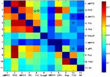
          Method: To reveal specific subsystem changes, we calculated the strength and number of FCS in the DMN intra- and inter-subsystems across individuals and compared the FC of the two groups. To further examine which pairs of brain regional functional connections contributed to the subsystem alterations, correlation coefficients between any two brain regions in the DMN were compared across groups. Additionally, to identify which regions made the strongest contributions to the subsystem changes, we calculated the regional FC strength (FCS), which was compared across groups.

          Results: For the intra-subsystem, decreased FC number and strength occurred in the MTL subsystem of AD patients but not in the DMPFC subsystem or core. For the inter-subsystems, the AD group showed decreased FCS and number between the MTL subsystem and PCC and a decreased number between the PCC and DMPFC subsystem. Decreased inter-regional FCS were found within the MTL subsystem in AD patients relative to controls: The posterior inferior parietal lobule (pIPL) showed decreased FC with the hippocampal formation (HF), parahippocampal cortex (PHC) and ventral MPFC (vMPFC). Decreased inter-regional FCS of the inter-subsystems were also found in AD patients: The HF and/or PHC showed decreased FC with dMPFC and TPJ, located in the DMPFC subsystem, and with PCC. AD patients also showed decreased FC between the PCC and TLC of the dMPFC subsystem. Furthermore, the HF and PHC in the MTL subsystem showed decreased regional FCS.

          Conclusion: Decreased intrinsic FC was mainly associated with the MTL subsystem of the AD group, suggesting that the MTL subsystem is predominantly disrupted.

          Related collections

          Most cited references38

          • Record: found
          • Abstract: found
          • Article: not found

          Searching for a baseline: functional imaging and the resting human brain.

          Functional brain imaging in humans has revealed task-specific increases in brain activity that are associated with various mental activities. In the same studies, mysterious, task-independent decreases have also frequently been encountered, especially when the tasks of interest have been compared with a passive state, such as simple fixation or eyes closed. These decreases have raised the possibility that there might be a baseline or resting state of brain function involving a specific set of mental operations. We explore this possibility, including the manner in which we might define a baseline and the implications of such a baseline for our understanding of brain function.
            Bookmark
            • Record: found
            • Abstract: found
            • Article: not found

            Remembering the past and imagining the future: common and distinct neural substrates during event construction and elaboration.

            People can consciously re-experience past events and pre-experience possible future events. This fMRI study examined the neural regions mediating the construction and elaboration of past and future events. Participants were cued with a noun for 20s and instructed to construct a past or future event within a specified time period (week, year, 5-20 years). Once participants had the event in mind, they made a button press and for the remainder of the 20s elaborated on the event. Importantly, all events generated were episodic and did not differ on a number of phenomenological qualities (detail, emotionality, personal significance, field/observer perspective). Conjunction analyses indicated the left hippocampus was commonly engaged by past and future event construction, along with posterior visuospatial regions, but considerable neural differentiation was also observed during the construction phase. Future events recruited regions involved in prospective thinking and generation processes, specifically right frontopolar cortex and left ventrolateral prefrontal cortex, respectively. Furthermore, future event construction uniquely engaged the right hippocampus, possibly as a response to the novelty of these events. In contrast to the construction phase, elaboration was characterized by remarkable overlap in regions comprising the autobiographical memory retrieval network, attributable to the common processes engaged during elaboration, including self-referential processing, contextual and episodic imagery. This striking neural overlap is consistent with findings that amnesic patients exhibit deficits in both past and future thinking, and confirms that the episodic system contributes importantly to imagining the future.
              Bookmark
              • Record: found
              • Abstract: found
              • Article: not found

              The functional neuroanatomy of autobiographical memory: a meta-analysis.

              Autobiographical memory (AM) entails a complex set of operations, including episodic memory, self-reflection, emotion, visual imagery, attention, executive functions, and semantic processes. The heterogeneous nature of AM poses significant challenges in capturing its behavioral and neuroanatomical correlates. Investigators have recently turned their attention to the functional neuroanatomy of AM. We used the effect-location method of meta-analysis to analyze data from 24 functional imaging studies of AM. The results indicated a core neural network of left-lateralized regions, including the medial and ventrolateral prefrontal, medial and lateral temporal and retrosplenial/posterior cingulate cortices, the temporoparietal junction and the cerebellum. Secondary and tertiary regions, less frequently reported in imaging studies of AM, are also identified. We examined the neural correlates of putative component processes in AM, including, executive functions, self-reflection, episodic remembering and visuospatial processing. We also separately analyzed the effect of select variables on the AM network across individual studies, including memory age, qualitative factors (personal significance, level of detail and vividness), semantic and emotional content, and the effect of reference conditions. We found that memory age effects on medial temporal lobe structures may be modulated by qualitative aspects of memory. Studies using rest as a control task masked process-specific components of the AM neural network. Our findings support a neural distinction between episodic and semantic memory in AM. Finally, emotional events produced a shift in lateralization of the AM network with activation observed in emotion-centered regions and deactivation (or lack of activation) observed in regions associated with cognitive processes.
                Bookmark

                Author and article information

                Contributors
                Journal
                Front Aging Neurosci
                Front Aging Neurosci
                Front. Aging Neurosci.
                Frontiers in Aging Neuroscience
                Frontiers Media S.A.
                1663-4365
                31 October 2018
                2018
                : 10
                : 344
                Affiliations
                [1] 1Department of Medical Imaging, Tongji Hospital, Tongji University School of Medicine, Tongji University , Shanghai, China
                [2] 2Department of Imaging, Huashan Hospital, Fudan University , Shanghai, China
                [3] 3Department of Imaging, The Fifth People’s Hospital of Shanghai, Fudan University , Shanghai, China
                [4] 4Department of Imaging, Shanghai Tongji Hospital , Shanghai, China
                Author notes

                Edited by: David Baglietto-Vargas, University of California, Irvine, United States

                Reviewed by: Aaron Wilber, Florida State University, United States; Manuel Francisco Lopez-Aranda, University of California, Los Angeles, United States; Alina Stimmell, Florida State University, United States, in collaboration with reviewer AW

                *Correspondence: Xiaohu Zhao, xhzhao999@ 123456263.net
                Article
                10.3389/fnagi.2018.00344
                6220065
                a27a1241-5753-42f6-a255-1e2956953388
                Copyright © 2018 Qi, Liu, Hu, He and Zhao.

                This is an open-access article distributed under the terms of the Creative Commons Attribution License (CC BY). The use, distribution or reproduction in other forums is permitted, provided the original author(s) and the copyright owner(s) are credited and that the original publication in this journal is cited, in accordance with accepted academic practice. No use, distribution or reproduction is permitted which does not comply with these terms.

                History
                : 10 July 2018
                : 11 October 2018
                Page count
                Figures: 5, Tables: 2, Equations: 0, References: 63, Pages: 10, Words: 0
                Categories
                Neuroscience
                Original Research

                Neurosciences
                resting-state fmri,default network,subsystems,functional connectivity,alzheimer’s disease

                Comments

                Comment on this article