13
views
0
recommends
+1 Recommend
0 collections
    0
    shares
      • Record: found
      • Abstract: found
      • Article: found
      Is Open Access

      Super-resolution microscopy reveals ultra-low CD19 expression on myeloma cells that triggers elimination by CD19 CAR-T

      research-article

      Read this article at

      Bookmark
          There is no author summary for this article yet. Authors can add summaries to their articles on ScienceOpen to make them more accessible to a non-specialist audience.

          Abstract

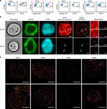
          Immunotherapy with chimeric antigen receptor-engineered T-cells (CAR-T) is under investigation in multiple myeloma. There are reports of myeloma remission after CD19 CAR-T therapy, although CD19 is hardly detectable on myeloma cells by flow cytometry (FC). We apply single molecule-sensitive direct stochastic optical reconstruction microscopy ( dSTORM), and demonstrate CD19 expression on a fraction of myeloma cells (10.3–80%) in 10 out of 14 patients (density: 13–5,000 molecules per cell). In contrast, FC detects CD19 in only 2 of these 10 patients, on a smaller fraction of cells. Treatment with CD19 CAR-T in vitro results in elimination of CD19-positive myeloma cells, including those with <100 CD19 molecules per cell. Similar data are obtained by dSTORM analyses of CD20 expression on myeloma cells and CD20 CAR-T. These data establish a sensitivity threshold for CAR-T and illustrate how super-resolution microscopy can guide patient selection in immunotherapy to exploit ultra-low density antigens.

          Abstract

          CD19 CAR-T cells have achieved some success in treating myeloma patients despite the limited detection of the CD19 antigen. Here, the authors show using dSTORM that 10/14 myeloma samples studied express ultra-low levels of CD19, which are sufficient for engaging CAR-T cells in vitro.

          Related collections

          Most cited references15

          • Record: found
          • Abstract: found
          • Article: not found

          CD22-CAR T Cells Induce Remissions in CD19-CAR Naïve and Resistant B-ALL

          Chimeric antigen receptor (CAR) T-cells targeting CD19 mediate potent effects in relapsed/refractory pre-B cell acute lymphoblastic leukemia (B-ALL) but antigen loss is a frequent cause of resistance to CD19-targeted immunotherapy. CD22 is also expressed on most B-ALL and usually retained following CD19 loss. We report results from a phase I trial testing a novel CD22-CAR in twenty-one children and adults, including 17 previously treated with CD19-directed immunotherapy. Dose dependent anti-leukemic activity was observed with complete remission in 73% (11/15) of patients receiving ≥ 1 × 10⁶ CD22-CART cells/kg, including 5/5 patients with CD19dim/neg B-ALL. Median remission duration was 6 months. Relapses were associated with diminished CD22 site density that likely permitted escape from killing by CD22-CART cells. These results are the first to eastablish the clinical activity of a CD22-CAR in pre-B cell ALL, including in leukemia resistant to anti-CD19 immunotherapy, demonstrating comparable potency to CD19-CART at biologically active doses in B-ALL. They also highlight the critical role played by antigen density in regulating CAR function. (Funded by NCI Intramural Research Program)
            Bookmark
            • Record: found
            • Abstract: found
            • Article: not found

            Direct stochastic optical reconstruction microscopy with standard fluorescent probes.

            Direct stochastic optical reconstruction microscopy (dSTORM) uses conventional fluorescent probes such as labeled antibodies or chemical tags for subdiffraction resolution fluorescence imaging with a lateral resolution of ∼20 nm. In contrast to photoactivated localization microscopy (PALM) with photoactivatable fluorescent proteins, dSTORM experiments start with bright fluorescent samples in which the fluorophores have to be transferred to a stable and reversible OFF state. The OFF state has a lifetime in the range of 100 milliseconds to several seconds after irradiation with light intensities low enough to ensure minimal photodestruction. Either spontaneously or photoinduced on irradiation with a second laser wavelength, a sparse subset of fluorophores is reactivated and their positions are precisely determined. Repetitive activation, localization and deactivation allow a temporal separation of spatially unresolved structures in a reconstructed image. Here we present a step-by-step protocol for dSTORM imaging in fixed and living cells on a wide-field fluorescence microscope, with standard fluorescent probes focusing especially on the photoinduced fine adjustment of the ratio of fluorophores residing in the ON and OFF states. Furthermore, we discuss labeling strategies, acquisition parameters, and temporal and spatial resolution. The ultimate step of data acquisition and data processing can be performed in seconds to minutes.
              Bookmark
              • Record: found
              • Abstract: found
              • Article: not found

              A transgene-encoded cell surface polypeptide for selection, in vivo tracking, and ablation of engineered cells.

              An unmet need in cell engineering is the availability of a single transgene encoded, functionally inert, human polypeptide that can serve multiple purposes, including ex vivo cell selection, in vivo cell tracking, and as a target for in vivo cell ablation. Here we describe a truncated human EGFR polypeptide (huEGFRt) that is devoid of extracellular N-terminal ligand binding domains and intracellular receptor tyrosine kinase activity but retains the native amino acid sequence, type I transmembrane cell surface localization, and a conformationally intact binding epitope for pharmaceutical-grade anti-EGFR monoclonal antibody, cetuximab (Erbitux). After lentiviral transduction of human T cells with vectors that coordinately express tumor-specific chimeric antigen receptors and huEGFRt, we show that huEGFRt serves as a highly efficient selection epitope for chimeric antigen receptor(+) T cells using biotinylated cetuximab in conjunction with current good manufacturing practices (cGMP)-grade anti-biotin immunomagnetic microbeads. Moreover, huEGFRt provides a cell surface marker for in vivo tracking of adoptively transferred T cells using both flow cytometry and immunohistochemistry, and a target for cetuximab-mediated antibody-dependent cellular cytotoxicity and in vivo elimination. The versatility of huEGFRt and the availability of pharmaceutical-grade reagents for its clinical application denote huEGFRt as a significant new tool for cellular engineering.
                Bookmark

                Author and article information

                Contributors
                m.sauer@uni-wuerzburg.de
                Hudecek_M@ukw.de
                Journal
                Nat Commun
                Nat Commun
                Nature Communications
                Nature Publishing Group UK (London )
                2041-1723
                17 July 2019
                17 July 2019
                2019
                : 10
                : 3137
                Affiliations
                [1 ]ISNI 0000 0001 1378 7891, GRID grid.411760.5, Medizinische Klinik und Poliklinik II, , Universitätsklinikum Würzburg, ; Oberdürrbacherstrasse 6, 97080 Würzburg, Germany
                [2 ]ISNI 0000 0001 1958 8658, GRID grid.8379.5, Department of Biotechnology and Biophysics, , Biocenter, Julius Maximilian University Würzburg, Am Hubland, ; 97074 Würzburg, Germany
                Author information
                http://orcid.org/0000-0002-0493-1369
                http://orcid.org/0000-0002-6983-1182
                http://orcid.org/0000-0003-1316-0241
                Article
                10948
                10.1038/s41467-019-10948-w
                6637169
                31316055
                a525b258-1c1f-4b05-9e5e-50ed3e2e98c3
                © The Author(s) 2019

                Open Access This article is licensed under a Creative Commons Attribution 4.0 International License, which permits use, sharing, adaptation, distribution and reproduction in any medium or format, as long as you give appropriate credit to the original author(s) and the source, provide a link to the Creative Commons license, and indicate if changes were made. The images or other third party material in this article are included in the article’s Creative Commons license, unless indicated otherwise in a credit line to the material. If material is not included in the article’s Creative Commons license and your intended use is not permitted by statutory regulation or exceeds the permitted use, you will need to obtain permission directly from the copyright holder. To view a copy of this license, visit http://creativecommons.org/licenses/by/4.0/.

                History
                : 24 March 2019
                : 10 June 2019
                Funding
                Funded by: This work was supported by the project “Center for Personalized Molecular Immunotherapy” funded within the EFRE-program of the European Union (Europäischer Fond für regionale Entwicklung); a grant from the German Ministry for Science and Education (BMBF, Bundesministerium für Bildung und Forschung, Grant # 13N14414); and a grant within the Max Eder Program of German Cancer Aid (Deutsche Krebshilfe e.V., Grant number 110313 to M.H.). M.H. is a member of the Young Scholar Program (Junges Kolleg) and Extraordinary Member of the Bavarian Academy of Sciences (Bayerische Akademie der Wissenschaften).
                Categories
                Article
                Custom metadata
                © The Author(s) 2019

                Uncategorized
                cancer imaging,cancer immunotherapy,imaging
                Uncategorized
                cancer imaging, cancer immunotherapy, imaging

                Comments

                Comment on this article