16
views
0
recommends
+1 Recommend
0 collections
    0
    shares
      • Record: found
      • Abstract: found
      • Article: found
      Is Open Access

      A Comprehensive Guide for Performing Sample Preparation and Top-Down Protein Analysis

      review-article

      Read this article at

      Bookmark
          There is no author summary for this article yet. Authors can add summaries to their articles on ScienceOpen to make them more accessible to a non-specialist audience.

          Abstract

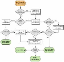
          Methodologies for the global analysis of proteins in a sample, or proteome analysis, have been available since 1975 when Patrick O′Farrell published the first paper describing two-dimensional gel electrophoresis (2D-PAGE). This technique allowed the resolution of single protein isoforms, or proteoforms, into single ‘spots’ in a polyacrylamide gel, allowing the quantitation of changes in a proteoform′s abundance to ascertain changes in an organism′s phenotype when conditions change. In pursuit of the comprehensive profiling of the proteome, significant advances in technology have made the identification and quantitation of intact proteoforms from complex mixtures of proteins more routine, allowing analysis of the proteome from the ‘Top-Down’. However, the number of proteoforms detected by Top-Down methodologies such as 2D-PAGE or mass spectrometry has not significantly increased since O’Farrell’s paper when compared to Bottom-Up, peptide-centric techniques. This article explores and explains the numerous methodologies and technologies available to analyse the proteome from the Top-Down with a strong emphasis on the necessity to analyse intact proteoforms as a better indicator of changes in biology and phenotype. We arrive at the conclusion that the complete and comprehensive profiling of an organism′s proteome is still, at present, beyond our reach but the continuing evolution of protein fractionation techniques and mass spectrometry brings comprehensive Top-Down proteome profiling closer.

          Related collections

          Most cited references182

          • Record: found
          • Abstract: found
          • Article: not found

          Stable isotope labeling by amino acids in cell culture, SILAC, as a simple and accurate approach to expression proteomics.

          Quantitative proteomics has traditionally been performed by two-dimensional gel electrophoresis, but recently, mass spectrometric methods based on stable isotope quantitation have shown great promise for the simultaneous and automated identification and quantitation of complex protein mixtures. Here we describe a method, termed SILAC, for stable isotope labeling by amino acids in cell culture, for the in vivo incorporation of specific amino acids into all mammalian proteins. Mammalian cell lines are grown in media lacking a standard essential amino acid but supplemented with a non-radioactive, isotopically labeled form of that amino acid, in this case deuterated leucine (Leu-d3). We find that growth of cells maintained in these media is no different from growth in normal media as evidenced by cell morphology, doubling time, and ability to differentiate. Complete incorporation of Leu-d3 occurred after five doublings in the cell lines and proteins studied. Protein populations from experimental and control samples are mixed directly after harvesting, and mass spectrometric identification is straightforward as every leucine-containing peptide incorporates either all normal leucine or all Leu-d3. We have applied this technique to the relative quantitation of changes in protein expression during the process of muscle cell differentiation. Proteins that were found to be up-regulated during this process include glyceraldehyde-3-phosphate dehydrogenase, fibronectin, and pyruvate kinase M2. SILAC is a simple, inexpensive, and accurate procedure that can be used as a quantitative proteomic approach in any cell culture system.
            Bookmark
            • Record: found
            • Abstract: found
            • Article: not found

            Quantitative analysis of complex protein mixtures using isotope-coded affinity tags.

            We describe an approach for the accurate quantification and concurrent sequence identification of the individual proteins within complex mixtures. The method is based on a class of new chemical reagents termed isotope-coded affinity tags (ICATs) and tandem mass spectrometry. Using this strategy, we compared protein expression in the yeast Saccharomyces cerevisiae, using either ethanol or galactose as a carbon source. The measured differences in protein expression correlated with known yeast metabolic function under glucose-repressed conditions. The method is redundant if multiple cysteinyl residues are present, and the relative quantification is highly accurate because it is based on stable isotope dilution techniques. The ICAT approach should provide a widely applicable means to compare quantitatively global protein expression in cells and tissues.
              Bookmark
              • Record: found
              • Abstract: found
              • Article: not found

              High resolution two-dimensional electrophoresis of proteins.

              A technique has been developed for the separation of proteins by two-dimensional polyacrylamide gel electrophoresis. Due to its resolution and sensitivity, this technique is a powerful tool for the analysis and detection of proteins from complex biological sources. Proteins are separated according to isoelectric point by isoelectric focusing in the first dimension, and according to molecular weight by sodium dodecyl sulfate electrophoresis in the second dimension. Since these two parameters are unrelated, it is possible to obtain an almost uniform distribution of protein spots across a two-diminsional gel. This technique has resolved 1100 different components from Escherichia coli and should be capable of resolving a maximum of 5000 proteins. A protein containing as little as one disintegration per min of either 14C or 35S can be detected by autoradiography. A protein which constitutes 10 minus 4 to 10 minus 5% of the total protein can be detected and quantified by autoradiography. The reproducibility of the separation is sufficient to permit each spot on one separation to be matched with a spot on a different separation. This technique provides a method for estimation (at the described sensitivities) of the number of proteins made by any biological system. This system can resolve proteins differing in a single charge and consequently can be used in the analysis of in vivo modifications resulting in a change in charge. Proteins whose charge is changed by missense mutations can be identified. A detailed description of the methods as well as the characteristics of this system are presented.
                Bookmark

                Author and article information

                Contributors
                Role: Academic Editor
                Role: Academic Editor
                Role: Academic Editor
                Journal
                Proteomes
                Proteomes
                proteomes
                Proteomes
                MDPI
                2227-7382
                07 April 2017
                June 2017
                : 5
                : 2
                : 11
                Affiliations
                [1 ]Proteomics Core Facility, University of Technology Sydney, PO Box 123, Broadway, Ultimo NSW 2007, Australia; iain.j.berry@ 123456student.uts.edu.au (I.J.B.); matthew.orourke@ 123456sydney.edu.au (M.B.O.R.); benjamin.raymond@ 123456uts.edu.au (B.B.A.R.); jerran.santos@ 123456uts.edu.au (J.S.); steven.djordjevic@ 123456uts.edu.au (S.P.D.)
                [2 ]Infection, Immunity and Innovation Institute, University of Technology Sydney, PO Box 123, Broadway, Ultimo NSW 2007, Australia
                [3 ]Mass Spectrometry Core Facility, Charles Perkins Centre, University of Sydney, The Hub D17, Sydney, Camperdown NSW 2006, Australia
                [4 ]Advanced Tissue Regeneration & Drug Delivery Group, University of Technology Sydney, PO Box 123, Broadway, Ultimo NSW 2007, Australia
                Author notes
                [* ]Correspondence: matthew.padula@ 123456uts.edu.au ; Tel.: +61-2-9514-4019
                Article
                proteomes-05-00011
                10.3390/proteomes5020011
                5489772
                28387712
                ad6f2fcf-ece9-4f07-a891-16bd367e8cc0
                © 2017 by the authors.

                Licensee MDPI, Basel, Switzerland. This article is an open access article distributed under the terms and conditions of the Creative Commons Attribution (CC BY) license ( http://creativecommons.org/licenses/by/4.0/).

                History
                : 26 December 2016
                : 04 April 2017
                Categories
                Review

                proteomics,top-down,bottom-up,mass spectrometry,electrophoresis,isoelectric focusing,proteoform,chromatography

                Comments

                Comment on this article