10
views
0
recommends
+1 Recommend
0 collections
    0
    shares
      • Record: found
      • Abstract: found
      • Article: found
      Is Open Access

      Energetics of discrete selectivity bands and mutation-induced transitions in the calcium-sodium ion channels family

      Preprint

      Read this article at

      Bookmark
          There is no author summary for this article yet. Authors can add summaries to their articles on ScienceOpen to make them more accessible to a non-specialist audience.

          Abstract

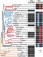
          We use Brownian dynamics simulations to study the permeation properties of a generic electrostatic model of a biological ion channel as a function of the fixed charge \(Q_f\) at its selectivity filter. We reconcile the recently-discovered discrete calcium conduction bands M0 (\(Q_f\)=1e), M1 (3e), M2 (5e) with the set of sodium conduction bands L0 (0.5-0.7e), L1 (1.5-2e) thereby obtaining a completed pattern of conduction and selectivity bands v \(Q_f\) for the sodium-calcium channels family. An increase of \(Q_f\) leads to an increase of calcium selectivity: L0 (sodium selective, non-blocking channel) -> M0 (non-selective channel) -> L1 (sodium selective channel with divalent block) -> M1 (calcium selective channel exhibiting the anomalous mole fraction effect). We create a consistent identification scheme where the L0 band is identified with the eukaryotic (DEKA) sodium channel, and L1/L2 (speculatively) with the bacterial NaChBac channel. The scheme created is able to account for the experimentally observed mutation-induced transformations between non-selective channels, sodium-selective channels, and calcium-selective channels, which we interpret as transitions between different rows of the identification table. By considering the potential energy changes during permeation, we show explicitly that the multi-ion conduction bands of calcium and sodium channels arise as the result of resonant barrier-less conduction. Our results confirm the crucial influence of electrostatic interactions on conduction and on the Ca/Na valence selectivity of calcium and sodium ion channels. The model and results could be also applicable to biomimetic nanopores with charged walls.

          Related collections

          Most cited references7

          • Record: found
          • Abstract: not found
          • Article: not found

          Mechanism of ion permeation through calcium channels

            Bookmark
            • Record: found
            • Abstract: found
            • Article: not found

            Probing ion-channel pores one proton at a time.

            Although membrane proteins often rely on ionizable residues for structure and function, their ionization states under physiological conditions largely elude experimental estimation. To gain insight into the effect of the local microenvironment on the proton affinity of ionizable residues, we have engineered individual lysines, histidines and arginines along the alpha-helical lining of the transmembrane pore of the nicotinic acetylcholine receptor. We can detect individual proton binding-unbinding reactions electrophysiologically at the level of a single proton on a single side chain as brief blocking-unblocking events of the passing cation current. Kinetic analysis of these fluctuations yields the position-dependent rates of proton transfer, from which the corresponding pK(a) values and shifts in pK(a) can be calculated. Here we present a self-consistent, residue-by-residue description of the microenvironment around the pore-lining transmembrane alpha-helices (M2) in the open-channel conformation, in terms of the excess free energy that is required to keep the engineered basic side chains protonated relative to bulk water. A comparison with closed-channel data leads us to propose that the rotation of M2, which is frequently invoked as a hallmark of the gating mechanism of Cys-loop receptors, is minimal, if any.
              Bookmark
              • Record: found
              • Abstract: found
              • Article: found
              Is Open Access

              NALCN Ion Channels Have Alternative Selectivity Filters Resembling Calcium Channels or Sodium Channels

              NALCN is a member of the family of ion channels with four homologous, repeat domains that include voltage-gated calcium and sodium channels. NALCN is a highly conserved gene from simple, extant multicellular organisms without nervous systems such as sponges and placozoans and mostly remains a single gene compared to the calcium and sodium channels which diversified into twenty genes in humans. The single NALCN gene has alternatively-spliced exons at exons 15 or exon 31 that splices in novel selectivity filter residues that resemble calcium channels (EEEE) or sodium channels (EKEE or EEKE). NALCN channels with alternative calcium, (EEEE) and sodium, (EKEE or EEKE) -selective pores are conserved in simple bilaterally symmetrical animals like flatworms to non-chordate deuterostomes. The single NALCN gene is limited as a sodium channel with a lysine (K)-containing pore in vertebrates, but originally NALCN was a calcium-like channel, and evolved to operate as both a calcium channel and sodium channel for different roles in many invertebrates. Expression patterns of NALCN-EKEE in pond snail, Lymnaea stagnalis suggest roles for NALCN in secretion, with an abundant expression in brain, and an up-regulation in secretory organs of sexually-mature adults such as albumen gland and prostate. NALCN-EEEE is equally abundant as NALCN-EKEE in snails, but is greater expressed in heart and other muscle tissue, and 50% less expressed in the brain than NALCN-EKEE. Transfected snail NALCN-EEEE and NALCN-EKEE channel isoforms express in HEK-293T cells. We were not able to distinguish potential NALCN currents from background, non-selective leak conductances in HEK293T cells. Native leak currents without expressing NALCN genes in HEK-293T cells are NMDG+ impermeant and blockable with 10 µM Gd3+ ions and are indistinguishable from the hallmark currents ascribed to mammalian NALCN currents expressed in vitro by Lu et al. in Cell. 2007 Apr 20;129(2):371-83.
                Bookmark

                Author and article information

                Journal
                08 May 2013
                2013-11-24
                Article
                10.1103/PhysRevE.88.052712
                1305.1847
                aec0a776-b10c-4bd4-ae7d-f4a6aa0b4fd6

                http://arxiv.org/licenses/nonexclusive-distrib/1.0/

                History
                Custom metadata
                Phys. Rev. E 88, 052712 (2013)
                16 pages, 17 figures, 2 tables, 75 references
                physics.bio-ph q-bio.BM

                Molecular biology,Biophysics
                Molecular biology, Biophysics

                Comments

                Comment on this article