34
views
0
recommends
+1 Recommend
0 collections
    0
    shares
      • Record: found
      • Abstract: found
      • Article: found
      Is Open Access

      Role of TRPM2 in H 2O 2-Induced Cell Apoptosis in Endothelial Cells

      research-article

      Read this article at

      Bookmark
          There is no author summary for this article yet. Authors can add summaries to their articles on ScienceOpen to make them more accessible to a non-specialist audience.

          Abstract

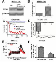
          Melastatin-like transient receptor potential channel 2 (TRPM2) is an oxidant-sensitive and cationic non-selective channel that is expressed in mammalian vascular endothelium. Here we investigated the functional role of TRPM2 channels in hydrogen peroxide (H 2O 2)-induced cytosolic Ca 2+ ([Ca 2+] i) elavation, whole-cell current increase, and apoptotic cell death in murine heart microvessel endothelial cell line H5V. A TRPM2 blocking antibody (TM2E3), which targets the E3 region near the ion permeation pore of TRPM2, was developed. Treatment of H5V cells with TM2E3 reduced the [Ca 2+] i rise and whole-cell current change in response to H 2O 2. Suppressing TRPM2 expression using TRPM2-specific short hairpin RNA (shRNA) had similar inhibitory effect. H 2O 2-induced apoptotic cell death in H5V cells was examined using MTT assay, DNA ladder formation analysis, and DAPI-based nuclear DNA condensation assay. Based on these assays, TM2E3 and TRPM2-specific shRNA both showed protective effect against H 2O 2-induced apoptotic cell death. TM2E3 and TRPM2-specific shRNA also protect the cells from tumor necrosis factor (TNF)-α-induced cell death in MTT assay. In contrast, overexpression of TRPM2 in H5V cells resulted in an increased response in [Ca 2+] i and whole-cell currents to H 2O 2. TRPM2 overexpression also aggravated the H 2O 2-induced apoptotic cell death. Downstream pathways following TRPM2 activation was examined. Results showed that TRPM2 activity stimulated caspase-8, caspase-9 and caspase-3. These findings strongly suggest that TRPM2 channel mediates cellular Ca 2+ overload in response to H 2O 2 and contribute to oxidant-induced apoptotic cell death in vascular endothelial cells. Down-regulating endogenous TRPM2 could be a means to protect the vascular endothelial cells from apoptotic cell death.

          Related collections

          Most cited references36

          • Record: found
          • Abstract: found
          • Article: not found

          ADP-ribose gating of the calcium-permeable LTRPC2 channel revealed by Nudix motif homology.

          Free ADP-ribose (ADPR), a product of NAD hydrolysis and a breakdown product of the calcium-release second messenger cyclic ADPR (cADPR), has no defined role as an intracellular signalling molecule in vertebrate systems. Here we show that a 350-amino-acid protein (designated NUDT9) and a homologous domain (NUDT9 homology domain) near the carboxy terminus of the LTRPC2/TrpC7 putative cation channel both function as specific ADPR pyrophosphatases. Whole-cell and single-channel analysis of HEK-293 cells expressing LTRPC2 show that LTRPC2 functions as a calcium-permeable cation channel that is specifically gated by free ADPR. The expression of native LTRPC2 transcripts is detectable in many tissues including the U937 monocyte cell line, in which ADPR induces large cation currents (designated IADPR) that closely match those mediated by recombinant LTRPC2. These results indicate that intracellular ADPR regulates calcium entry into cells that express LTRPC2.
            Bookmark
            • Record: found
            • Abstract: found
            • Article: not found

            Endothelial dysfunction: a multifaceted disorder (The Wiggers Award Lecture).

            Endothelial cells synthesize and release various factors that regulate angiogenesis, inflammatory responses, hemostasis, as well as vascular tone and permeability. Endothelial dysfunction has been associated with a number of pathophysiological processes. Oxidative stress appears to be a common denominator underlying endothelial dysfunction in cardiovascular diseases. However, depending on the pathology, the vascular bed studied, the stimulant, and additional factors such as age, sex, salt intake, cholesterolemia, glycemia, and hyperhomocysteinemia, the mechanisms underlying the endothelial dysfunction can be markedly different. A reduced bioavailability of nitric oxide (NO), an alteration in the production of prostanoids, including prostacyclin, thromboxane A2, and/or isoprostanes, an impairment of endothelium-dependent hyperpolarization, as well as an increased release of endothelin-1, can individually or in association contribute to endothelial dysfunction. Therapeutic interventions do not necessarily restore a proper endothelial function and, when they do, may improve only part of these variables.
              Bookmark
              • Record: found
              • Abstract: found
              • Article: not found

              Nitric oxide activates TRP channels by cysteine S-nitrosylation.

              Transient receptor potential (TRP) proteins form plasma-membrane cation channels that act as sensors for diverse cellular stimuli. Here, we report a novel activation mechanism mediated by cysteine S-nitrosylation in TRP channels. Recombinant TRPC1, TRPC4, TRPC5, TRPV1, TRPV3 and TRPV4 of the TRPC and TRPV families, which are commonly classified as receptor-activated channels and thermosensor channels, induce entry of Ca(2+) into cells in response to nitric oxide (NO). Labeling and functional assays using cysteine mutants, together with membrane sidedness in activating reactive disulfides, show that cytoplasmically accessible Cys553 and nearby Cys558 are nitrosylation sites mediating NO sensitivity in TRPC5. The responsive TRP proteins have conserved cysteines on the same N-terminal side of the pore region. Notably, nitrosylation of native TRPC5 upon G protein-coupled ATP receptor stimulation elicits entry of Ca(2+) into endothelial cells. These findings reveal the structural motif for the NO-sensitive activation gate in TRP channels and indicate that NO sensors are a new functional category of cellular receptors extending over different TRP families.
                Bookmark

                Author and article information

                Contributors
                Role: Editor
                Journal
                PLoS One
                PLoS ONE
                plos
                plosone
                PLoS ONE
                Public Library of Science (San Francisco, USA )
                1932-6203
                2012
                20 August 2012
                : 7
                : 8
                : e43186
                Affiliations
                [1 ]Li Ka Shing Institute of Health Sciences and School of Biomedical Sciences, The Chinese University of Hong Kong, Hong Kong, China
                [2 ]Stem Cell and Regenerative Medicine Consortium, The University of Hong Kong, Hong Kong, China
                Catholic University Medical School, Italy
                Author notes

                Competing Interests: The authors have declared that no competing interests exist.

                Conceived and designed the experiments: XQY. Performed the experiments: LS WYW. Analyzed the data: LS HYY WYW. Contributed reagents/materials/analysis tools: XQY RAL YH. Wrote the paper: LS XQY RAL. Edited and revised the manuscript: XQY RAL YH.

                Article
                PONE-D-12-01964
                10.1371/journal.pone.0043186
                3423428
                22916222
                b0a0c68e-d4f9-4ebd-8ce3-159dd59e7757
                Copyright @ 2012

                This is an open-access article distributed under the terms of the Creative Commons Attribution License, which permits unrestricted use, distribution, and reproduction in any medium, provided the original author and source are credited.

                History
                : 19 January 2012
                : 18 July 2012
                Page count
                Pages: 10
                Funding
                This work was supported by research grants CUHK477408, CUHK479109, and CUHK478710 from Hong Kong RGC, Strategic Investment Scheme C and Group Research Grant from Chinese University of Hong Kong, and National Science Foundation of China grant 31171100. The funders had no role in study design, data collection and analysis, decision to publish, or preparation of the manuscript.
                Categories
                Research Article
                Biology
                Anatomy and Physiology
                Cell Physiology
                Genetics
                Molecular Genetics
                Gene Regulation
                Molecular Cell Biology
                Cellular Types
                Endothelial Cells
                Cell Death
                Gene Expression
                Medicine
                Cardiovascular
                Vascular Biology
                Oncology
                Cancer Treatment
                Antibody Therapy

                Uncategorized
                Uncategorized

                Comments

                Comment on this article