3
views
0
recommends
+1 Recommend
0 collections
    0
    shares
      • Record: found
      • Abstract: found
      • Article: found
      Is Open Access

      Liver structural transformation after partial hepatectomy and repeated partial hepatectomy in rats: A renewed view on liver regeneration

      research-article

      Read this article at

      Bookmark
          There is no author summary for this article yet. Authors can add summaries to their articles on ScienceOpen to make them more accessible to a non-specialist audience.

          Abstract

          BACKGROUND

          The phenomenon of liver regeneration after partial hepatectomy (PH) is still a subject of considerable interest due to the increasing frequency of half liver transplantation on the one hand, and on the other hand, new surgical approaches which allow removal of massive space-occupying hepatic tumors, which earlier was considered as inoperable. Interestingly, the mechanisms of liver regeneration are extensively studied after PH but less attention is paid to the architectonics of the regenerated organ. Because of this, the question “How does the structure of regenerated liver differ from normal, regular liver?” has not been fully answered yet. Furthermore, almost without any attention is left the liver's structural transformation after repeated hepatectomy (of the re-regenereted liver).

          AIM

          To compare the architectonics of the lobules and circulatory bed of normal, re-generated and re-regenerated livers.

          METHODS

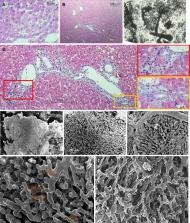
          The livers of 40 adult, male, albino Wistar rats were studied. 14 rats were subjected to PH - the 1 st study group (SG1); 10 rats underwent repeated PH – the 2 nd study group (SG2); 16 rats were subjected to sham operation - control group (CG); The livers were studied after 9 months from PH, and after 6 months from repeated PH. Cytological (Schiff reaction for the determination of DNA concen-tration), histological (H&E, Masson trichrome, CK8 Immunohistochemical marker, transparent slides after Indian Ink injection, ), morphometrical (hepatocytes areas, perimeters and ploidy) and Electron Microscopical (Scanning Electron Microscopy of corrosion casts) methods were used.

          RESULTS

          In the SG1 and SG2, the area of hepatocytes and their perimeter are increased compared to the CG ( P < 0.05). However, the areas and perimeters of the hepatocytes of the SG 1 and SG 2 groups reveal a lesser difference. In regenerated (SG 1) and re-regenerated (SG 2) livers, the hepatocytes form the remodeled lobules, which size (300-1200 µm) exceeds the sizes of the lobules from CG (300-600 µm). The remodeled lobules (especially the “mega-lobules” with the sizes 1000-1200 µm) contain the transformed meshworks of the sinusoids, the part of which is dilated asymmetrically. This meshwork might have originated from the several portal venules (interlobular and/or inlet). The boundaries between the adjacent lobules (including mega-lobules) are widened and filled by connective tissue fibers, which gives the liver parenchyma a nodular look. In SG 2 the unevenness of sinusoid diameters, as well as the boundaries between the lobules (including the mega-lobules) are more vividly expressed in comparison with SG 1. The liver tissue of both SG 1 and SG 2 is featured by the slightly expressed ductular reaction.

          CONCLUSION

          Regenerated and re-regenerated livers in comparison with normal liver contain hypertrophied hepatocytes with increased ploidy which together with transformed sinusoidal and biliary meshworks form the remodeled lobulli.

          Related collections

          Most cited references53

          • Record: found
          • Abstract: found
          • Article: not found

          Liver regeneration.

          Liver regeneration after partial hepatectomy is a very complex and well-orchestrated phenomenon. It is carried out by the participation of all mature liver cell types. The process is associated with signaling cascades involving growth factors, cytokines, matrix remodeling, and several feedbacks of stimulation and inhibition of growth related signals. Liver manages to restore any lost mass and adjust its size to that of the organism, while at the same time providing full support for body homeostasis during the entire regenerative process. In situations when hepatocytes or biliary cells are blocked from regeneration, these cell types can function as facultative stem cells for each other.
            Bookmark
            • Record: found
            • Abstract: found
            • Article: not found

            Liver regeneration.

            Liver regeneration after the loss of hepatic tissue is a fundamental parameter of liver response to injury. Recognized as a phenomenon from mythological times, it is now defined as an orchestrated response induced by specific external stimuli and involving sequential changes in gene expression, growth factor production, and morphologic structure. Many growth factors and cytokines, most notably hepatocyte growth factor, epidermal growth factor, transforming growth factor-alpha, interleukin-6, tumor necrosis factor-alpha, insulin, and norepinephrine, appear to play important roles in this process. This review attempts to integrate the findings of the last three decades and looks toward clues as to the nature of the causes that trigger this fascinating organ and cellular response.
              Bookmark
              • Record: found
              • Abstract: found
              • Article: not found

              Dynamics of endothelial cell behavior in sprouting angiogenesis.

              The vertebrate body contains an extensive blood vessel network that forms, with a few exceptions, by endothelial sprouting from the existing vasculature. This process, termed angiogenesis, involves complex and highly dynamic interactions between endothelial cells and their environment. Pro-angiogenic signals, such as VEGF, promote endothelial motility, filopodia extension and proliferation, and, together with Notch signaling, controls whether specific endothelial cells become lead tip cells or trailing stalk cells. Sprouts then convert into endothelial tubules and form connections with other vessels, which requires the local suppression of motility and the formation of new cell-cell junctions. We here review the dynamics of angiogenesis in the context of key molecules and pathways controlling tip cell selection, sprouting and the formation of new vessels. Copyright © 2010 Elsevier Ltd. All rights reserved.
                Bookmark

                Author and article information

                Contributors
                Journal
                World J Gastroenterol
                World J. Gastroenterol
                WJG
                World Journal of Gastroenterology
                Baishideng Publishing Group Inc
                1007-9327
                2219-2840
                21 July 2020
                21 July 2020
                : 26
                : 27
                : 3899-3916
                Affiliations
                Faculty of Medicine, Ivane Javakhishvili Tbilisi State University, Tbilisi 0159, Georgia
                Faculty of Medicine, Ivane Javakhishvili Tbilisi State University, Tbilisi 0159, Georgia
                Faculty of Medicine, Ivane Javakhishvili Tbilisi State University, Tbilisi 0159, Georgia
                Faculty of Medicine, Ivane Javakhishvili Tbilisi State University, Tbilisi 0159, Georgia
                Faculty of Medicine, Ivane Javakhishvili Tbilisi State University, Tbilisi 0159, Georgia
                Faculty of Exact and Natural Sciences, Ivane Javakhishvili Tbilisi State University, Tbilisi 0179, Georgia
                Faculty of Exact and Natural Sciences, Ivane Javakhishvili Tbilisi State University, Tbilisi 0179, Georgia
                Department of Surgical Oncology (Surgery C), Chaim Sheba Medical Center at HaShomer, Tel Aviv 52621, Israel
                Federal Research and Clinical Center of Physical-Chemical Medicine of Federal Medical Biological Agency, Moscow 119435, Russia
                Faculty of Medicine, Ivane Javakhishvili Tbilisi State University, Tbilisi 0159, Georgia
                Clinical Anatomy and Operative Surgery, Ivane Javakhishvili Tbilisi State University, Tbilisi 0159, Georgia. dimitri.kordzaia@ 123456tsu.ge
                Author notes

                Author contributions: Tsomaia K and Patarashvili L were responsible for planing experiments, the preparation and evaluation of histological slides, for mor-phometrical, literature search, in data interpretation and manuscript drafting; Karumidze N and Modebadze I were responsible for determination of the DNA concentration of the hepatocytes; Bebiashvili I and Azmaipharashvili E contributed in experiments, in literature search and review and in analysis of the results. Dzidziguri D and Sareli M contributed in planning the experiments and research methodology, in data analysis, manuscript drafting and revision; Gusev S performed SEM investigation, evaluation, description, data analysis, editing and revision of the manuscript; Kordzaia D performed the conceptualization of research, project administration, supervision of experiments and investigation of SEM. Control preparation of the last version of the manuscript; all authors reviewed and approved the final version to be published.

                Supported by the Shota Rustaveli National Science Foundation of Georgia, No. DP2016_22 [New Interfaculty Interdisciplinary Structured Doctoral Programme “ Translational Biomedicine” (Direction – “ Hepatology” )].

                Corresponding author: Dimitri Kordzaia, DSc, MD, PhD, Dean, Professor, Clinical Anatomy and Operative Surgery, Ivane Javakhishvili Tbilisi State University, Beliashvili str. 78, Tbilisi 0159, Georgia. dimitri.kordzaia@ 123456tsu.ge

                Article
                jWJG.v26.i27.pg3899
                10.3748/wjg.v26.i27.3899
                7385567
                32774065
                b1036d7f-86c3-4c5a-b160-eef00b56139b
                ©The Author(s) 2020. Published by Baishideng Publishing Group Inc. All rights reserved.

                This article is an open-access article which was selected by an in-house editor and fully peer-reviewed by external reviewers. It is distributed in accordance with the Creative Commons Attribution Non Commercial (CC BY-NC 4.0) license, which permits others to distribute, remix, adapt, build upon this work non-commercially, and license their derivative works on different terms, provided the original work is properly cited and the use is non-commercial.

                History
                : 27 February 2020
                : 12 May 2020
                : 23 June 2020
                Categories
                Basic Study

                partial hepatectomy,repeated hepatectomy,liver regeneration,liver re-regeneration,hepatocytes hypertrophy,remodeling,ploidy,corrosion casts

                Comments

                Comment on this article