55
views
0
recommends
+1 Recommend
0 collections
    0
    shares
      • Record: found
      • Abstract: found
      • Article: found
      Is Open Access

      The DDN Catalytic Motif Is Required for Metnase Functions in Non-homologous End Joining (NHEJ) Repair and Replication Restart*

      research-article

      Read this article at

      Bookmark
          There is no author summary for this article yet. Authors can add summaries to their articles on ScienceOpen to make them more accessible to a non-specialist audience.

          Abstract

          Background: Metnase, a transposase-containing DNA repair protein, retains DNA cleavage activity with a DDN motif.

          Results: Substitution with the ancestral transposase DDD/DDE catalytic motif results in a decrease in ssDNA binding and ss-overhang cleavage activities.

          Conclusion: The DDN motif is required for Metnase DNA repair activities.

          Significance: Understanding the requirements for catalytic activity provides insights on how Metnase functions as a DNA repair protein.

          Abstract

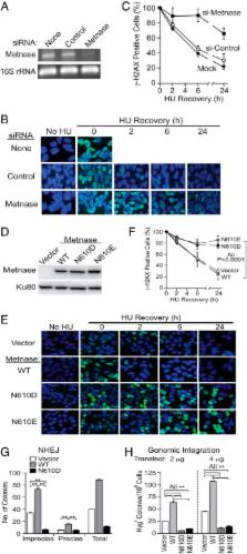
          Metnase (or SETMAR) arose from a chimeric fusion of the Hsmar1 transposase downstream of a protein methylase in anthropoid primates. Although the Metnase transposase domain has been largely conserved, its catalytic motif (DDN) differs from the DDD motif of related transposases, which may be important for its role as a DNA repair factor and its enzymatic activities. Here, we show that substitution of DDN 610 with either DDD 610 or DDE 610 significantly reduced in vivo functions of Metnase in NHEJ repair and accelerated restart of replication forks. We next tested whether the DDD or DDE mutants cleave single-strand extensions and flaps in partial duplex DNA and pseudo-Tyr structures that mimic stalled replication forks. Neither substrate is cleaved by the DDD or DDE mutant, under the conditions where wild-type Metnase effectively cleaves ssDNA overhangs. We then characterized the ssDNA-binding activity of the Metnase transposase domain and found that the catalytic domain binds ssDNA but not dsDNA, whereas dsDNA binding activity resides in the helix-turn-helix DNA binding domain. Substitution of Asn-610 with either Asp or Glu within the transposase domain significantly reduces ssDNA binding activity. Collectively, our results suggest that a single mutation DDN 610 → DDD 610, which restores the ancestral catalytic site, results in loss of function in Metnase.

          Related collections

          Most cited references32

          • Record: found
          • Abstract: found
          • Article: not found

          Ulp1-SUMO crystal structure and genetic analysis reveal conserved interactions and a regulatory element essential for cell growth in yeast.

          Modification of cellular proteins by the ubiquitin-like protein SUMO is essential for nuclear processes and cell cycle progression in yeast. The Ulp1 protease catalyzes two essential functions in the SUMO pathway: (1) processing of full-length SUMO to its mature form and (2) deconjugation of SUMO from targeted proteins. Selective reduction of the proteolytic reaction produced a covalent thiohemiacetal transition state complex between a Ulp1 C-terminal fragment and its cellular substrate Smt3, the yeast SUMO homolog. The Ulp1-Smt3 crystal structure and functional testing of elements within the conserved interface elucidate determinants of SUMO recognition, processing, and deconjugation. Genetic analysis guided by the structure further reveals a regulatory element N-terminal to the proteolytic domain that is required for cell growth in yeast.
            Bookmark
            • Record: found
            • Abstract: found
            • Article: not found

            Transposition mediated by RAG1 and RAG2 and its implications for the evolution of the immune system.

            Immunoglobulin and T-cell-receptor genes are assembled from component gene segments in developing lymphocytes by a site-specific recombination reaction, V(D)J recombination. The proteins encoded by the recombination-activating genes, RAG1 and RAG2, are essential in this reaction, mediating sequence-specific DNA recognition of well-defined recombination signals and DNA cleavage next to these signals. Here we show that RAG1 and RAG2 together form a transposase capable of excising a piece of DNA containing recombination signals from a donor site and inserting it into a target DNA molecule. The products formed contain a short duplication of target DNA immediately flanking the transposed fragment, a structure like that created by retroviral integration and all known transposition reactions. The results support the theory that RAG1 and RAG2 were once components of a transposable element, and that the split nature of immunoglobulin and T-cell-receptor genes derives from germline insertion of this element into an ancestral receptor gene soon after the evolutionary divergence of jawed and jawless vertebrates.
              Bookmark
              • Record: found
              • Abstract: found
              • Article: not found

              The evolutionary history of human DNA transposons: evidence for intense activity in the primate lineage.

              Class 2, or DNA transposons, make up approximately 3% of the human genome, yet the evolutionary history of these elements has been largely overlooked and remains poorly understood. Here we carried out the first comprehensive analysis of the activity of human DNA transposons over the course of primate evolution using three independent computational methods. First, we conducted an exhaustive search for human DNA transposons nested within L1 and Alu elements known to be primate specific. Second, we assessed the presence/absence of 794 human DNA transposons at orthologous positions in 10 mammalian species using sequence data generated by The ENCODE Project. These two approaches, which do not rely upon sequence divergence, allowed us to classify DNA transposons into three different categories: anthropoid specific (40-63 My), primate specific (64-80 My), and eutherian wide (81-150 My). Finally, we used this data to calculate the substitution rates of DNA transposons for each category and refine the age of each family based on the average percent divergence of individual copies to their consensus. Based on these combined methods, we can confidently estimate that at least 40 human DNA transposon families, representing approximately 98,000 elements ( approximately 33 Mb) in the human genome, have been active in the primate lineage. There was a cessation in the transpositional activity of DNA transposons during the later phase of the primate radiation, with no evidence of elements younger than approximately 37 My. This data points to intense activity of DNA transposons during the mammalian radiation and early primate evolution, followed, apparently, by their mass extinction in an anthropoid primate ancestor.
                Bookmark

                Author and article information

                Journal
                J Biol Chem
                J. Biol. Chem
                jbc
                jbc
                JBC
                The Journal of Biological Chemistry
                American Society for Biochemistry and Molecular Biology (9650 Rockville Pike, Bethesda, MD 20814, U.S.A. )
                0021-9258
                1083-351X
                11 April 2014
                25 February 2014
                25 February 2014
                : 289
                : 15
                : 10930-10938
                Affiliations
                From the []Department of Biochemistry and Molecular Biology, Indiana University School of Medicine, Indianapolis, Indiana 46202,
                the [§ ]Department of Environmental and Radiological Health Sciences, Colorado State University, Fort Collins, Colorado 80523,
                the []Department of Medicine, University of Florida and Shands Health Care System, Gainesville, Florida 32610, and
                the []Department of Chemistry and Chemical Biology, Indiana University Purdue University Indianapolis, Indianapolis, Indiana 46202
                Author notes
                [1 ] To whom correspondence should be addressed: Dept. of Biochemistry and Molecular Biology, Indiana University Simon Cancer Center, Indiana University School of Medicine, Indianapolis, IN. Tel.: 317-278-3464; Fax: 317-274-8046; E-mail: slee@ 123456iu.edu .
                Article
                M113.533216
                10.1074/jbc.M113.533216
                4036204
                24573677
                b35bff5d-4c01-40d3-99be-4e094c38a00e
                © 2014 by The American Society for Biochemistry and Molecular Biology, Inc.

                Author's Choice—Final version full access.

                Creative Commons Attribution Unported License applies to Author Choice Articles

                History
                : 6 November 2013
                : 11 February 2014
                Funding
                Funded by: National Institutes of Health
                Award ID: R01 CA151367
                Award ID: R01 GM084020
                Award ID: R01 CA140422
                Categories
                DNA and Chromosomes

                Biochemistry
                dna binding protein,dna damage,dna enzymes,dna repair,dna replication
                Biochemistry
                dna binding protein, dna damage, dna enzymes, dna repair, dna replication

                Comments

                Comment on this article