22
views
0
recommends
+1 Recommend
0 collections
    0
    shares
      • Record: found
      • Abstract: found
      • Article: found
      Is Open Access

      Functional hypoxia drives neuroplasticity and neurogenesis via brain erythropoietin

      research-article

      Read this article at

      Bookmark
          There is no author summary for this article yet. Authors can add summaries to their articles on ScienceOpen to make them more accessible to a non-specialist audience.

          Abstract

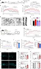
          Erythropoietin (EPO), named after its role in hematopoiesis, is also expressed in mammalian brain. In clinical settings, recombinant EPO treatment has revealed a remarkable improvement of cognition, but underlying mechanisms have remained obscure. Here, we show with a novel line of reporter mice that cognitive challenge induces local/endogenous hypoxia in hippocampal pyramidal neurons, hence enhancing expression of EPO and EPO receptor (EPOR). High-dose EPO administration, amplifying auto/paracrine EPO/EPOR signaling, prompts the emergence of new CA1 neurons and enhanced dendritic spine densities. Single-cell sequencing reveals rapid increase in newly differentiating neurons. Importantly, improved performance on complex running wheels after EPO is imitated by exposure to mild exogenous/inspiratory hypoxia. All these effects depend on neuronal expression of the Epor gene. This suggests a model of neuroplasticity in form of a fundamental regulatory circle, in which neuronal networks—challenged by cognitive tasks—drift into transient hypoxia, thereby triggering neuronal EPO/EPOR expression.

          Abstract

          EPO treatment improves cognition, but underlying mechanisms were unknown. Here the authors describe a regulatory loop in which brain networks challenged by cognitive tasks drift into functional hypoxia that drives—via neuronal EPO synthesis—neurodifferentiation and dendritic spine formation.

          Related collections

          Most cited references42

          • Record: found
          • Abstract: found
          • Article: not found

          Transcriptional regulation of vascular endothelial cell responses to hypoxia by HIF-1.

          Hypoxia-inducible factor 1 (HIF-1) activates transcription of genes encoding angiogenic growth factors, which are secreted by hypoxic cells and stimulate endothelial cells, leading to angiogenesis. To determine whether HIF-1 also mediates cell-autonomous responses to hypoxia, we have compared gene expression profiles in arterial endothelial cells cultured under nonhypoxic versus hypoxic conditions and in nonhypoxic cells infected with adenovirus encoding beta-galactosidase versus a constitutively active form of HIF-1alpha (AdCA5). There were 245 gene probes that showed at least 1.5-fold increase in expression in response to hypoxia and in response to AdCA5; 325 gene probes showed at least 1.5-fold decrease in expression in response to hypoxia and in response to AdCA5. The largest category of genes down-regulated by both hypoxia and AdCA5 encoded proteins involved in cell growth/proliferation. Many genes up-regulated by both hypoxia and AdCA5 encoded cytokines/growth factors, receptors, and other signaling proteins. Transcription factors accounted for the largest group of HIF-1-regulated genes, indicating that HIF-1 controls a network of transcriptional responses to hypoxia in endothelial cells. Infection of endothelial cells with AdCA5 under nonhypoxic conditions was sufficient to induce increased basement membrane invasion and tube formation similar to the responses induced by hypoxia, indicating that HIF-1 mediates cell-autonomous activation of endothelial cells.
            Bookmark
            • Record: found
            • Abstract: found
            • Article: not found

            Corridors of Migrating Neurons in Human Brain and Their Decline during Infancy

            The subventricular zone (SVZ) of many adult non-human mammals generates large numbers of new neurons destined for the olfactory bulb (OB) 1–6 . Along the walls of the lateral ventricles, immature neuronal progeny migrate in tangentially-oriented chains that coalesce into a rostral migratory stream (RMS) connecting the SVZ to the OB. The adult human SVZ, in contrast, contains a hypocellular gap layer separating the ependymal lining from a periventricular ribbon of astrocytes 7 . Some of these SVZ astrocytes can function as neural stem cells in vitro, but their function in vivo remains controversial. An initial report finds few SVZ proliferating cells and rare migrating immature neurons in the RMS of adult humans 7 . In contrast, a subsequent study indicates robust proliferation and migration in the human SVZ and RMS 8,9 . Here, we find that the infant human SVZ and RMS contain an extensive corridor of migrating immature neurons before 18 months of age, but, contrary to previous reports 8 , this germinal activity subsides in older children and is nearly extinct by adulthood. Surprisingly, during this limited window of neurogenesis, not all new neurons in the human SVZ are destined for the OB – we describe a major migratory pathway that targets the prefrontal cortex in humans. Together, these findings reveal robust streams of tangentially migrating immature neurons in human early postnatal SVZ and cortex. These pathways represent potential targets of neurological injuries affecting neonates.
              Bookmark
              • Record: found
              • Abstract: found
              • Article: not found

              Structural dynamics of dendritic spines in memory and cognition.

              Recent studies show that dendritic spines are dynamic structures. Their rapid creation, destruction and shape-changing are essential for short- and long-term plasticity at excitatory synapses on pyramidal neurons in the cerebral cortex. The onset of long-term potentiation, spine-volume growth and an increase in receptor trafficking are coincident, enabling a 'functional readout' of spine structure that links the age, size, strength and lifetime of a synapse. Spine dynamics are also implicated in long-term memory and cognition: intrinsic fluctuations in volume can explain synapse maintenance over long periods, and rapid, activity-triggered plasticity can relate directly to cognitive processes. Thus, spine dynamics are cellular phenomena with important implications for cognition and memory. Furthermore, impaired spine dynamics can cause psychiatric and neurodevelopmental disorders. Copyright 2010 Elsevier Ltd. All rights reserved.
                Bookmark

                Author and article information

                Contributors
                nave@em.mpg.de
                ehrenreich@em.mpg.de
                Journal
                Nat Commun
                Nat Commun
                Nature Communications
                Nature Publishing Group UK (London )
                2041-1723
                9 March 2020
                9 March 2020
                2020
                : 11
                : 1313
                Affiliations
                [1 ]ISNI 0000 0001 0668 6902, GRID grid.419522.9, Clinical Neuroscience, , Max Planck Institute of Experimental Medicine, ; Göttingen, Germany
                [2 ]ISNI 0000 0001 2173 938X, GRID grid.5338.d, Neurobiology Unit, Program in Neurosciences and Interdisciplinary Research Structure for Biotechnology and Biomedicine (BIOTECMED), , Universitat de València, ; Burjassot, Spain
                [3 ]ISNI 0000 0001 2180 3484, GRID grid.13648.38, Institute of Medical Systems Biology, Center for Molecular Neurobiology, , University Clinic Hamburg-Eppendorf, ; Hamburg, Germany
                [4 ]ISNI 0000 0001 0668 6902, GRID grid.419522.9, Department of Neurogenetics, , Max Planck Institute of Experimental Medicine, ; Göttingen, Germany
                [5 ]Synaptic Systems GmbH, Göttingen, Germany
                [6 ]GRID grid.475435.4, Copenhagen Affective Disorder Research Centre, Psychiatric Centre Copenhagen, , Copenhagen University Hospital, Rigshospitalet, ; Copenhagen, Denmark
                [7 ]ISNI 0000 0001 0668 6902, GRID grid.419522.9, Department of Molecular Neurobiology, , Max Planck Institute of Experimental Medicine, ; Göttingen, Germany
                [8 ]DFG Research Center for Nanoscale Microscopy and Molecular Physiology of the Brain (CNMPB), Göttingen, Germany
                [9 ]CIBERSAM: Spanish National Network for Research in Mental Health, Valencia, Spain
                [10 ]GRID grid.411308.f, Fundación Investigación Hospital Clínico de Valencia, INCLIVA, ; Valencia, Spain
                Author information
                http://orcid.org/0000-0003-3288-857X
                http://orcid.org/0000-0001-9112-4444
                http://orcid.org/0000-0001-8087-360X
                http://orcid.org/0000-0002-0944-7226
                http://orcid.org/0000-0001-7913-2716
                http://orcid.org/0000-0002-6741-8861
                http://orcid.org/0000-0002-7083-1358
                http://orcid.org/0000-0002-4222-0947
                http://orcid.org/0000-0002-7908-7369
                http://orcid.org/0000-0003-3452-8500
                http://orcid.org/0000-0002-7710-5738
                http://orcid.org/0000-0002-5589-983X
                http://orcid.org/0000-0003-2572-1384
                http://orcid.org/0000-0001-8724-9666
                http://orcid.org/0000-0001-8371-5711
                Article
                15041
                10.1038/s41467-020-15041-1
                7062779
                32152318
                b3a3eff1-cd7c-4a9d-a660-572d55ffe713
                © The Author(s) 2020

                Open Access This article is licensed under a Creative Commons Attribution 4.0 International License, which permits use, sharing, adaptation, distribution and reproduction in any medium or format, as long as you give appropriate credit to the original author(s) and the source, provide a link to the Creative Commons license, and indicate if changes were made. The images or other third party material in this article are included in the article’s Creative Commons license, unless indicated otherwise in a credit line to the material. If material is not included in the article’s Creative Commons license and your intended use is not permitted by statutory regulation or exceeds the permitted use, you will need to obtain permission directly from the copyright holder. To view a copy of this license, visit http://creativecommons.org/licenses/by/4.0/.

                History
                : 15 October 2019
                : 12 February 2020
                Funding
                Funded by: National University of Sciences and Technology (NUST), Faculty Development Program Abroad 2014/15 Pakistan
                Funded by: IMPRS-GGNB Ph.D. Program Neurosciences (DFG Grant GSC 226) at the Georg-August-University
                Funded by: FundRef https://doi.org/10.13039/100005984, Dr. Miriam and Sheldon G. Adelson Medical Research Foundation (Dr. Miriam & Sheldon G. Adelson Medical Research Foundation);
                Funded by: ERC Advanced Grant
                Funded by: FundRef https://doi.org/10.13039/501100004189, Max-Planck-Gesellschaft (Max Planck Society);
                Funded by: DFG Research Center for Nanoscale Microscopy and Molecular Physiology of the Brain (CNMPB)
                Categories
                Article
                Custom metadata
                © The Author(s) 2020

                Uncategorized
                cellular neuroscience,cognitive neuroscience
                Uncategorized
                cellular neuroscience, cognitive neuroscience

                Comments

                Comment on this article