2
views
0
recommends
+1 Recommend
0 collections
    0
    shares
      • Record: found
      • Abstract: found
      • Article: found
      Is Open Access

      Dynamic contrast-enhanced MRI radiomics nomogram for predicting axillary lymph node metastasis in breast cancer

      research-article

      Read this article at

      Bookmark
          There is no author summary for this article yet. Authors can add summaries to their articles on ScienceOpen to make them more accessible to a non-specialist audience.

          Abstract

          Purpose

          The goal of this study is to develop and validate a radiomics nomogram integrating the radiomics features from DCE-MRI and clinical factors for the preoperative diagnosis of axillary lymph node (ALN) metastasis in breast cancer patients.

          Procedures

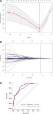
          A total of 432 patients with breast cancer were enrolled in this retrospective study and divided into a training cohort ( n = 296) and a validation cohort ( n = 136). Radiomics features were extracted from the second phase of dynamic contrast enhanced (DCE) MRI images. The least absolute shrinkage and selection operator (LASSO) regression method was used to screen optimal features and construct a radiomics signature in the training cohort. Multivariable logistic regression analysis was used to establish a radiomics nomogram model based on the radiomics signature and clinical factors. The predictive performance of the nomogram was quantified with respect to discrimination and calibration, which was further evaluated in the independent validation cohort.

          Results

          Fourteen ALN metastasis-related features were selected to construct the radiomics signature, with an area under the curve (AUC) of 0.847 and 0.805 in the training and validation cohorts, respectively. The nomogram was established by incorporating the histological grade, multifocality, MRI report lymph node status and radiomics signature and showed good calibration and excellent performance for ALN detection (AUC of 0.907 and 0.874 in the training and validation cohorts, respectively). The decision curve, which demonstrated the radiomics nomogram, displayed promising clinical utility.

          Conclusions

          The radiomics nomogram can be used as a noninvasive and reliable tool to assist clinicians in accurately predicting ALN metastasis in breast cancer preoperatively.

          Supplementary Information

          The online version contains supplementary material available at 10.1186/s40644-022-00450-w.

          Related collections

          Most cited references43

          • Record: found
          • Abstract: found
          • Article: not found

          Cancer incidence and mortality worldwide: sources, methods and major patterns in GLOBOCAN 2012.

          Estimates of the worldwide incidence and mortality from 27 major cancers and for all cancers combined for 2012 are now available in the GLOBOCAN series of the International Agency for Research on Cancer. We review the sources and methods used in compiling the national cancer incidence and mortality estimates, and briefly describe the key results by cancer site and in 20 large "areas" of the world. Overall, there were 14.1 million new cases and 8.2 million deaths in 2012. The most commonly diagnosed cancers were lung (1.82 million), breast (1.67 million), and colorectal (1.36 million); the most common causes of cancer death were lung cancer (1.6 million deaths), liver cancer (745,000 deaths), and stomach cancer (723,000 deaths). © 2014 UICC.
            Bookmark
            • Record: found
            • Abstract: found
            • Article: not found

            Radiomics: Images Are More than Pictures, They Are Data

            This report describes the process of radiomics, its challenges, and its potential power to facilitate better clinical decision making, particularly in the care of patients with cancer.
              Bookmark
              • Record: found
              • Abstract: found
              • Article: not found

              Radiomics: the bridge between medical imaging and personalized medicine

              Radiomics, the high-throughput mining of quantitative image features from standard-of-care medical imaging that enables data to be extracted and applied within clinical-decision support systems to improve diagnostic, prognostic, and predictive accuracy, is gaining importance in cancer research. Radiomic analysis exploits sophisticated image analysis tools and the rapid development and validation of medical imaging data that uses image-based signatures for precision diagnosis and treatment, providing a powerful tool in modern medicine. Herein, we describe the process of radiomics, its pitfalls, challenges, opportunities, and its capacity to improve clinical decision making, emphasizing the utility for patients with cancer. Currently, the field of radiomics lacks standardized evaluation of both the scientific integrity and the clinical relevance of the numerous published radiomics investigations resulting from the rapid growth of this area. Rigorous evaluation criteria and reporting guidelines need to be established in order for radiomics to mature as a discipline. Herein, we provide guidance for investigations to meet this urgent need in the field of radiomics.
                Bookmark

                Author and article information

                Contributors
                haisongdeling@126.com
                haiyangfei@126.com
                961934757@qq.com
                308545220@qq.com
                396908371@qq.com
                312563541@qq.com
                hbzjkzyx@163.com
                s312016581@163.com
                Journal
                Cancer Imaging
                Cancer Imaging
                Cancer Imaging
                BioMed Central (London )
                1740-5025
                1470-7330
                4 April 2022
                4 April 2022
                2022
                : 22
                : 17
                Affiliations
                [1 ]GRID grid.412026.3, ISNI 0000 0004 1776 2036, Graduate Faculty, , Hebei North University, ; 12 Changqing Road, Qiaoxi District, Zhangjiakou, 075000 China
                [2 ]GRID grid.414906.e, ISNI 0000 0004 1808 0918, Department of Radiology, , The First Affiliated Hospital of Wenzhou Medical University, ; Nanbaixiang New District, Ouhai District, Wenzhou, 32000 Zhejiang China
                [3 ]GRID grid.412026.3, ISNI 0000 0004 1776 2036, Department of Radiology, , The First Affiliated Hospital of Hebei North University, ; 12 Changqing Road, Qiaoxi District, Zhangjiakou, 075000 China
                Author information
                http://orcid.org/0000-0002-4640-302X
                Article
                450
                10.1186/s40644-022-00450-w
                8981871
                35379339
                b5296f59-3f73-49e0-9a4a-a3cd0177c880
                © The Author(s) 2022

                Open AccessThis article is licensed under a Creative Commons Attribution 4.0 International License, which permits use, sharing, adaptation, distribution and reproduction in any medium or format, as long as you give appropriate credit to the original author(s) and the source, provide a link to the Creative Commons licence, and indicate if changes were made. The images or other third party material in this article are included in the article's Creative Commons licence, unless indicated otherwise in a credit line to the material. If material is not included in the article's Creative Commons licence and your intended use is not permitted by statutory regulation or exceeds the permitted use, you will need to obtain permission directly from the copyright holder. To view a copy of this licence, visit http://creativecommons.org/licenses/by/4.0/. The Creative Commons Public Domain Dedication waiver ( http://creativecommons.org/publicdomain/zero/1.0/) applies to the data made available in this article, unless otherwise stated in a credit line to the data.

                History
                : 24 September 2020
                : 1 February 2022
                Funding
                Funded by: Graduate Research and Innovation Projects of Hebei Province
                Award ID: CXZZSS2021130
                Award Recipient :
                Categories
                Research Article
                Custom metadata
                © The Author(s) 2022

                breast cancer,axillary lymph node metastasis,radiomics,preoperative prediction

                Comments

                Comment on this article