6
views
0
recommends
+1 Recommend
0 collections
    0
    shares
      • Record: found
      • Abstract: found
      • Article: found
      Is Open Access

      Separability of Acute Cerebral Infarction Lesions in CT Based Radiomics: Toward Artificial Intelligence-Assisted Diagnosis

      research-article

      Read this article at

      Bookmark
          There is no author summary for this article yet. Authors can add summaries to their articles on ScienceOpen to make them more accessible to a non-specialist audience.

          Abstract

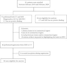
          This study aims at analyzing the separability of acute cerebral infarction lesions which were invisible in CT. 38 patients, who were diagnosed with acute cerebral infarction and performed both CT and MRI, and 18 patients, who had no positive finding in either CT or MRI, were enrolled. Comparative studies were performed on lesion and symmetrical regions, normal brain and symmetrical regions, lesion, and normal brain regions. MRI was reconstructed and affine transformed to obtain accurate lesion position of CT. Radiomic features and information gain were introduced to capture efficient features. Finally, 10 classifiers were established with selected features to evaluate the effectiveness of analysis. 1301 radiomic features were extracted from candidate regions after registration. For lesion and their symmetrical regions, there were 280 features with information gain greater than 0.1 and 2 features with information gain greater than 0.3. The average classification accuracy was 0.6467, and the best classification accuracy was 0.7748. For normal brain and their symmetrical regions, there were 176 features with information gain greater than 0.1, 1 feature with information gain greater than 0.2. The average classification accuracy was 0.5414, and the best classification accuracy was 0.6782. For normal brain and lesions, there were 501 features with information gain greater than 0.1 and 1 feature with information gain greater than 0.5. The average classification accuracy was 0.7480, and the best classification accuracy was 0.8694. In conclusion, the study captured significant features correlated with acute cerebral infarction and confirmed the separability of acute lesions in CT, which established foundation for further artificial intelligence-assisted CT diagnosis.

          Related collections

          Most cited references26

          • Record: found
          • Abstract: found
          • Article: not found

          Guidelines for the Early Management of Patients With Acute Ischemic Stroke: 2019 Update to the 2018 Guidelines for the Early Management of Acute Ischemic Stroke: A Guideline for Healthcare Professionals From the American Heart Association/American Stroke Association

          Background and Purpose- The purpose of these guidelines is to provide an up-to-date comprehensive set of recommendations in a single document for clinicians caring for adult patients with acute arterial ischemic stroke. The intended audiences are prehospital care providers, physicians, allied health professionals, and hospital administrators. These guidelines supersede the 2013 Acute Ischemic Stroke (AIS) Guidelines and are an update of the 2018 AIS Guidelines. Methods- Members of the writing group were appointed by the American Heart Association (AHA) Stroke Council's Scientific Statements Oversight Committee, representing various areas of medical expertise. Members were not allowed to participate in discussions or to vote on topics relevant to their relations with industry. An update of the 2013 AIS Guidelines was originally published in January 2018. This guideline was approved by the AHA Science Advisory and Coordinating Committee and the AHA Executive Committee. In April 2018, a revision to these guidelines, deleting some recommendations, was published online by the AHA. The writing group was asked review the original document and revise if appropriate. In June 2018, the writing group submitted a document with minor changes and with inclusion of important newly published randomized controlled trials with >100 participants and clinical outcomes at least 90 days after AIS. The document was sent to 14 peer reviewers. The writing group evaluated the peer reviewers' comments and revised when appropriate. The current final document was approved by all members of the writing group except when relationships with industry precluded members from voting and by the governing bodies of the AHA. These guidelines use the American College of Cardiology/AHA 2015 Class of Recommendations and Level of Evidence and the new AHA guidelines format. Results- These guidelines detail prehospital care, urgent and emergency evaluation and treatment with intravenous and intra-arterial therapies, and in-hospital management, including secondary prevention measures that are appropriately instituted within the first 2 weeks. The guidelines support the overarching concept of stroke systems of care in both the prehospital and hospital settings. Conclusions- These guidelines provide general recommendations based on the currently available evidence to guide clinicians caring for adult patients with acute arterial ischemic stroke. In many instances, however, only limited data exist demonstrating the urgent need for continued research on treatment of acute ischemic stroke.
            Bookmark
            • Record: found
            • Abstract: found
            • Article: not found

            Computational Radiomics System to Decode the Radiographic Phenotype

            Radiomics aims to quantify phenotypic characteristics on medical imaging through the use of automated algorithms. Radiomic artificial intelligence (AI) technology, either based on engineered hard-coded algorithms or deep learning methods, can be used to develop non-invasive imaging-based biomarkers. However, lack of standardized algorithm definitions and image processing severely hampers reproducibility and comparability of results. To address this issue, we developed PyRadiomics , a flexible open-source platform capable of extracting a large panel of engineered features from medical images. PyRadiomics is implemented in Python and can be used standalone or using 3D-Slicer. Here, we discuss the workflow and architecture of PyRadiomics and demonstrate its application in characterizing lung-lesions. Source code, documentation, and examples are publicly available at www.radiomics.io . With this platform, we aim to establish a reference standard for radiomic analyses, provide a tested and maintained resource, and to grow the community of radiomic developers addressing critical needs in cancer research.
              Bookmark
              • Record: found
              • Abstract: found
              • Article: not found

              Radiomics: extracting more information from medical images using advanced feature analysis.

              Solid cancers are spatially and temporally heterogeneous. This limits the use of invasive biopsy based molecular assays but gives huge potential for medical imaging, which has the ability to capture intra-tumoural heterogeneity in a non-invasive way. During the past decades, medical imaging innovations with new hardware, new imaging agents and standardised protocols, allows the field to move towards quantitative imaging. Therefore, also the development of automated and reproducible analysis methodologies to extract more information from image-based features is a requirement. Radiomics--the high-throughput extraction of large amounts of image features from radiographic images--addresses this problem and is one of the approaches that hold great promises but need further validation in multi-centric settings and in the laboratory. Copyright © 2011 Elsevier Ltd. All rights reserved.
                Bookmark

                Author and article information

                Contributors
                Journal
                Biomed Res Int
                Biomed Res Int
                BMRI
                BioMed Research International
                Hindawi
                2314-6133
                2314-6141
                2020
                15 November 2020
                : 2020
                : 8864756
                Affiliations
                1North University of China-Taiyuan Central Hospital Joint Innovation Institute, 3 Xueyuan Road, Taiyuan, Shanxi 030051, China
                2College of Big Data, North University of China, 3 Xueyuan Road, Taiyuan, Shanxi 030051, China
                3Taiyuan Central Hospital of Shanxi Medical University, 5 Dong San Dao Lane, Jiefang Street, Taiyuan, Shanxi 030009, China
                4School of Information and Communication Engineering, North University of China, 3 Xueyuan Road, Taiyuan, Shanxi 030051, China
                Author notes

                Academic Editor: Zhiguo Zhou

                Author information
                https://orcid.org/0000-0002-3941-5030
                https://orcid.org/0000-0003-2395-5961
                https://orcid.org/0000-0001-9099-9695
                https://orcid.org/0000-0002-8628-1266
                https://orcid.org/0000-0001-7755-7550
                https://orcid.org/0000-0002-9086-7366
                https://orcid.org/0000-0002-4992-1631
                Article
                10.1155/2020/8864756
                7683107
                b63f277b-4973-4870-9281-387e00095e3f
                Copyright © 2020 Yun Guan et al.

                This is an open access article distributed under the Creative Commons Attribution License, which permits unrestricted use, distribution, and reproduction in any medium, provided the original work is properly cited.

                History
                : 16 September 2020
                : 2 November 2020
                : 3 November 2020
                Funding
                Funded by: Science and Technology Foundation of North University of China
                Award ID: 20191634
                Funded by: Graduate Research and Innovation Projects of Shanxi Province
                Award ID: 2020SY381
                Funded by: Construction project of Engineering Technology Research Center of Shanxi Province
                Award ID: 201805D121008
                Funded by: project of Health Commission of Shanxi Province
                Award ID: 2018109
                Funded by: National Natural Science Foundation of China
                Award ID: 81671789
                Categories
                Research Article

                Comments

                Comment on this article