21
views
0
recommends
+1 Recommend
0 collections
    0
    shares
      • Record: found
      • Abstract: found
      • Article: found
      Is Open Access

      The effects of a genome-wide supported variant in the CACNA1C gene on cortical morphology in schizophrenia patients and healthy subjects

      research-article

      Read this article at

      Bookmark
          There is no author summary for this article yet. Authors can add summaries to their articles on ScienceOpen to make them more accessible to a non-specialist audience.

          Abstract

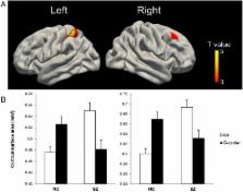
          Schizophrenia is a highly heritable disorder with multiple susceptibility genes. Previously, we identified CACNA1C rs2007044 as a new risk locus for schizophrenia, with the minor allele G as risk allele. This association was recently validated by a powerful genome-wide association study. However, the underlying neural mechanisms remain unclear. Therefore, we tested whether the risk allele has an influence on cortical surface area and thickness in a sample of schizophrenia patients and healthy controls. We found significant genotype by diagnosis interactions on cortical surface area, but not thickness, in the right dorsolateral prefrontal cortex and the left superior parietal cortex, both of which are key components of the central executive network. Moreover, the surface areas of both regions were inversely correlated with PANSS negative scores in AA homogeneous patients but not in G-carriers. This is the first study to describe the influence of the new genome-wide supported schizophrenia risk variant on cortical morphology. Our data revealed a significant genetic effect of cortical surface area in pivotal brain regions, which have been implicated in the pathophysiology of schizophrenia, possibly via their involvement in cognitive functions. These results yield new insights into the potential neural mechanisms linking CACNA1C to the risk of schizophrenia.

          Related collections

          Most cited references43

          • Record: found
          • Abstract: found
          • Article: not found

          A TALE nuclease architecture for efficient genome editing.

          Nucleases that cleave unique genomic sequences in living cells can be used for targeted gene editing and mutagenesis. Here we develop a strategy for generating such reagents based on transcription activator-like effector (TALE) proteins from Xanthomonas. We identify TALE truncation variants that efficiently cleave DNA when linked to the catalytic domain of FokI and use these nucleases to generate discrete edits or small deletions within endogenous human NTF3 and CCR5 genes at efficiencies of up to 25%. We further show that designed TALEs can regulate endogenous mammalian genes. These studies demonstrate the effective application of designed TALE transcription factors and nucleases for the targeted regulation and modification of endogenous genes.
            Bookmark
            • Record: found
            • Abstract: found
            • Article: not found

            Specification of cerebral cortical areas.

            P Rakic (1988)
            How the immense population of neurons that constitute the human cerebral neocortex is generated from progenitors lining the cerebral ventricle and then distributed to appropriate layers of distinctive cytoarchitectonic areas can be explained by the radial unit hypothesis. According to this hypothesis, the ependymal layer of the embryonic cerebral ventricle consists of proliferative units that provide a proto-map of prospective cytoarchitectonic areas. The output of the proliferative units is translated via glial guides to the expanding cortex in the form of ontogenetic columns, whose final number for each area can be modified through interaction with afferent input. Data obtained through various advanced neurobiological techniques, including electron microscopy, immunocytochemistry, [3H]thymidine and receptor autoradiography, retrovirus gene transfer, neural transplants, and surgical or genetic manipulation of cortical development, furnish new details about the kinetics of cell proliferation, their lineage relationships, and phenotypic expression that favor this hypothesis. The radial unit model provides a framework for understanding cerebral evolution, epigenetic regulation of the parcellation of cytoarchitectonic areas, and insight into the pathogenesis of certain cortical disorders in humans.
              Bookmark
              • Record: found
              • Abstract: found
              • Article: not found

              Automated manifold surgery: constructing geometrically accurate and topologically correct models of the human cerebral cortex.

              Highly accurate surface models of the cerebral cortex are becoming increasingly important as tools in the investigation of the functional organization of the human brain. The construction of such models is difficult using current neuroimaging technology due to the high degree of cortical folding. Even single voxel misclassifications can result in erroneous connections being created between adjacent banks of a sulcus, resulting in a topologically inaccurate model. These topological defects cause the cortical model to no longer be homeomorphic to a sheet, preventing the accurate inflation, flattening, or spherical morphing of the reconstructed cortex. Surface deformation techniques can guarantee the topological correctness of a model, but are time-consuming and may result in geometrically inaccurate models. In order to address this need we have developed a technique for taking a model of the cortex, detecting and fixing the topological defects while leaving that majority of the model intact, resulting in a surface that is both geometrically accurate and topologically correct.
                Bookmark

                Author and article information

                Journal
                Sci Rep
                Sci Rep
                Scientific Reports
                Nature Publishing Group
                2045-2322
                29 September 2016
                2016
                : 6
                : 34298
                Affiliations
                [1 ]Brainnetome Center, Institute of Automation, Chinese Academy of Sciences , Beijing, China
                [2 ]National Laboratory of Pattern Recognition, Institute of Automation, Chinese Academy of Sciences , Beijing, China
                [3 ]Institute of Mental Health, The Sixth Hospital, Peking University , Beijing, China
                [4 ]Key Laboratory of Mental Health, Ministry of Health & National Clinical Research Center for Mental Disorders (Peking University) , Beijing, China
                [5 ]Queensland Brain Institute, The University of Queensland, Brisbane , QLD, Australia
                [6 ]Key Laboratory for NeuroInformation of Ministry of Education, School of Life Science and Technology, University of Electronic Science and Technology of China , Chengdu, China
                Author notes
                [*]

                These authors contributed equally to this work.

                Article
                srep34298
                10.1038/srep34298
                5041147
                27683010
                bdef24c9-ad01-4817-97e1-eb834d5d9dd3
                Copyright © 2016, The Author(s)

                This work is licensed under a Creative Commons Attribution 4.0 International License. The images or other third party material in this article are included in the article’s Creative Commons license, unless indicated otherwise in the credit line; if the material is not included under the Creative Commons license, users will need to obtain permission from the license holder to reproduce the material. To view a copy of this license, visit http://creativecommons.org/licenses/by/4.0/

                History
                : 10 May 2016
                : 09 September 2016
                Categories
                Article

                Uncategorized
                Uncategorized

                Comments

                Comment on this article