10
views
0
recommends
+1 Recommend
0 collections
    0
    shares
      • Record: found
      • Abstract: found
      • Article: not found

      Mitigated NSAID-induced apoptotic and autophagic cell death with Smad7 overexpression

      research-article

      Read this article at

      ScienceOpenPublisherPMC
      Bookmark
          There is no author summary for this article yet. Authors can add summaries to their articles on ScienceOpen to make them more accessible to a non-specialist audience.

          Abstract

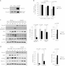
          Non-steroidal anti-inflammatory drugs damaged gastrointestinal mucosa in cyclooxygenase-dependent and -independent pathway, among which apopototic or autophagic cell death in gastrointestinal cells might be one of key cytotoxic mechanisms responsible for NSAID-induced damages. Therefore, alleviating this cell death after NSAIDs can be a rescuing strategy. In this study, we explored the role of Smad7 on NSAID-induced cytotoxicity in gastric epithelial cells. Using RGM1 cells, we have compared biological changes between mock-transfected and Smad7-overexpressed cells. As results, significantly decreased cytotoxicity accompanied with decreased levels of cleaved caspase-3 and poly (ADP-ribose) polymerase, Bax, and autophagic vesicles concurrent with decreased expressions of autophagy protein 5 and microtubule-associated protein light chain 3B-II were noted in Smad7-overexpressed cells with indomethacin administration compared to mock-transfected cells. Contrast to mitigated apoptotic execution, anti-apoptotic Bcl-2 and Beclin-1 were significantly increased in Smad7-overexpressed cells compared to mock-transfected cells. Smad7 siRNA significantly reversed these protective actions of Smad7 against indomethacin, in which p38 mitogen-activated protein kinase was significantly intervened. Furthermore, indomethacin-induced Smad7 degradation through ubiquitin-proteasome pathway was relevant to increased cytotoxicity, while chloroquine as autophagy inhibitor significantly attenuated indomethacin-induced cytotoxicity through Smad7 preservation via repressed ubiquitination. Conclusively, either genetic overexpression or pharmacological induction of Smad7 significantly attenuated indomethacin-induced gastric cell damages.

          Related collections

          Most cited references25

          • Record: found
          • Abstract: not found
          • Article: not found

          Inhibition of prostaglandin synthesis as a mechanism of action for aspirin-like drugs.

          J R Vane (1971)
            Bookmark
            • Record: found
            • Abstract: found
            • Article: not found

            Autophagy is activated for cell survival after endoplasmic reticulum stress.

            Eukaryotic cells deal with accumulation of unfolded proteins in the endoplasmic reticulum (ER) by the unfolded protein response, involving the induction of molecular chaperones, translational attenuation, and ER-associated degradation, to prevent cell death. Here, we found that the autophagy system is activated as a novel signaling pathway in response to ER stress. Treatment of SK-N-SH neuroblastoma cells with ER stressors markedly induced the formation of autophagosomes, which were recognized at the ultrastructural level. The formation of green fluorescent protein (GFP)-LC3-labeled structures (GFP-LC3 "dots"), representing autophagosomes, was extensively induced in cells exposed to ER stress with conversion from LC3-I to LC3-II. In IRE1-deficient cells or cells treated with c-Jun N-terminal kinase (JNK) inhibitor, the autophagy induced by ER stress was inhibited, indicating that the IRE1-JNK pathway is required for autophagy activation after ER stress. In contrast, PERK-deficient cells and ATF6 knockdown cells showed that autophagy was induced after ER stress in a manner similar to the wild-type cells. Disturbance of autophagy rendered cells vulnerable to ER stress, suggesting that autophagy plays important roles in cell survival after ER stress.
              Bookmark
              • Record: found
              • Abstract: found
              • Article: not found

              Endoplasmic reticulum stress signaling in disease.

              The extracellular space is an environment hostile to unmodified polypeptides. For this reason, many eukaryotic proteins destined for exposure to this environment through secretion or display at the cell surface require maturation steps within a specialized organelle, the endoplasmic reticulum (ER). A complex homeostatic mechanism, known as the unfolded protein response (UPR), has evolved to link the load of newly synthesized proteins with the capacity of the ER to mature them. It has become apparent that dysfunction of the UPR plays an important role in some human diseases, especially those involving tissues dedicated to extracellular protein synthesis. Diabetes mellitus is an example of such a disease, since the demands for constantly varying levels of insulin synthesis make pancreatic beta-cells dependent on efficient UPR signaling. Furthermore, recent discoveries in this field indicate that the importance of the UPR in diabetes is not restricted to the beta-cell but is also involved in peripheral insulin resistance. This review addresses aspects of the UPR currently understood to be involved in human disease, including their role in diabetes mellitus, atherosclerosis, and neoplasia.
                Bookmark

                Author and article information

                Journal
                J Clin Biochem Nutr
                J Clin Biochem Nutr
                JCBN
                Journal of Clinical Biochemistry and Nutrition
                the Society for Free Radical Research Japan (Kyoto, Japan )
                0912-0009
                1880-5086
                January 2017
                17 December 2016
                : 60
                : 1
                : 55-62
                Affiliations
                [1 ]Department of Biochemistry, Gachon University School of Medicine, 7-45, Songdo-dong, Yeonsu-ku, Incheon 406-840, Korea
                [2 ]CHA University Cancer Prevention Research Center, CHA Bio Complex, 335, Pangyo-ro, Bundang-gu, Seongnam 463-400, Korea
                [3 ]CHA University Bundang Medical Center, 59, Yatap-ro, Bundang-gu, Seongnam 463-712, Korea
                Author notes
                *To whom correspondence should be addressed. E-mail: hojlee@ 123456gachon.ac.kr (HJ Lee), hahmkb@ 123456cha.ac.kr (KB Hahm)
                Article
                jcbn16-69
                10.3164/jcbn.16-69
                5281534
                28163383
                be255a5d-58b1-4360-b312-5f9edc954573
                Copyright © 2017 JCBN

                This is an open access article distributed under the terms of the Creative Commons Attribution License, which permits unrestricted use, distribution, and reproduction in any medium, provided the original work is properly cited.

                History
                : 11 August 2016
                : 7 September 2016
                Categories
                Original Article

                Biochemistry
                nsaid,gastrointestinal damages,smad7,apoptosis,autophagy
                Biochemistry
                nsaid, gastrointestinal damages, smad7, apoptosis, autophagy

                Comments

                Comment on this article