296
views
0
recommends
+1 Recommend
0 collections
    8
    shares
      • Record: found
      • Abstract: found
      • Article: found
      Is Open Access

      In the Crosshairs: Investigating Lytic Granules by High-Resolution Microscopy and Electrophysiology

      review-article

      Read this article at

      Bookmark
          There is no author summary for this article yet. Authors can add summaries to their articles on ScienceOpen to make them more accessible to a non-specialist audience.

          Abstract

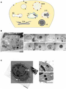
          Cytotoxic T lymphocytes (CTLs) form an integral part of the adaptive immune system. Their main function is to eliminate bacteria- and virus-infected target cells by releasing perforin and granzymes (the lethal hit) contained within lytic granules (LGs), at the CTL-target-cell interface [the immunological synapse (IS)]. The formation of the IS as well as the final events at the IS leading to target-cell death are both highly complex and dynamic processes. In this review we highlight and discuss three high-resolution techniques that have proven invaluable in the effort to decipher key features of the mechanism of CTL effector function and in particular lytic granule maturation and fusion. Correlative light and electron microscopy allows the correlation between organelle morphology and localization of particular proteins, while total internal reflection fluorescence microscopy (TIRFM) enables the study of lytic granule dynamics at the IS in real time. The combination of TIRFM with patch-clamp membrane capacitance measurements finally provides a tool to quantify the size of fusing LGs at the IS.

          Related collections

          Most cited references31

          • Record: found
          • Abstract: not found
          • Article: not found

          Beiträge zur Theorie des Mikroskops und der mikroskopischen Wahrnehmung

          E. Abbe (1873)
            Bookmark
            • Record: found
            • Abstract: found
            • Article: not found

            Protein localization in electron micrographs using fluorescence nanoscopy

            A complete portrait of a cell requires a detailed description of its molecular topography: proteins must be linked to particular organelles. Immuno-electron microscopy can reveal locations of proteins with nanometer resolution but is limited by the quality of fixation, the paucity of antibodies, and the inaccessibility of the antigens. Here, we describe correlative fluorescence electron microscopy for the nanoscopic localization of proteins in electron micrographs. Proteins tagged with Citrine or tdEos were expressed in Caenorhabditis elegans, fixed and embedded. Tagged proteins were imaged from ultrathin sections using stimulated emission depletion microscopy (STED) or photoactivated localization microscopy (PALM). Fluorescence was correlated with organelles imaged in electron micrographs from the same sections. These methods were used to successfully localize histones, a mitochondrial protein, and a presynaptic dense projection protein in electron micrographs.
              Bookmark
              • Record: found
              • Abstract: found
              • Article: not found

              Cytotoxic immunological synapses.

              One of the most fundamental activities of the adaptive immune system is to kill infected cells and tumor cells. Two distinct pathways mediate this process, both of which are facilitated by a cytotoxic immunological synapse. While traditionally thought of as innate immune cells, natural killer (NK) cells are now appreciated to have the capacity for long-term adaptation to chemical and viral insults. These cells integrate multiple positive and negative signals through NK cell cytotoxic or inhibitory synapses. The traditional CD8(+)alphabeta T-cell receptor-positive cells are among the best models for the concept of an immunological synapse, in which vectoral signaling is linked to directed secretion in a stable interface to induce apoptotic cell death in an infected cell. Large-scale molecular organization in synapses generated a number of hypotheses. Studies in the past 5 years have started to provide clear answers regarding the validity of these models. In vivo imaging approaches have provided some hints as to the physiologic relevance of these processes with great promise for the future. This review provides an overview of work on cytotoxic immunological synapses and suggests pathways forward in applying this information to the development of therapeutic agents.
                Bookmark

                Author and article information

                Contributors
                Journal
                Front Immunol
                Front Immunol
                Front. Immunol.
                Frontiers in Immunology
                Frontiers Media S.A.
                1664-3224
                27 November 2013
                2013
                : 4
                : 411
                Affiliations
                [1] 1Institute of Physiology, Saarland University , Homburg, Saar, Germany
                Author notes

                Edited by: Yenan Bryceson, Karolinska Institutet, Sweden

                Reviewed by: Subramaniam Malarkannan, Medical College of Wisconsin, USA; Clair Gardiner, Trinity College Dublin, Ireland

                *Correspondence: Jens Rettig, Physiologisches Institut, Universität des Saarlandes, Gebäude 59, Kirrberger Street 8, 66421 Homburg, Saar, Germany e-mail: jrettig@ 123456uks.eu

                This article was submitted to NK Cell Biology, a section of the journal Frontiers in Immunology.

                Article
                10.3389/fimmu.2013.00411
                3841761
                24348478
                bfc30850-981c-4076-979c-ecedff75c0fd
                Copyright © 2013 Pattu, Halimani, Ming, Schirra, Hahn, Bzeih, Chang, Weins, Krause and Rettig.

                This is an open-access article distributed under the terms of the Creative Commons Attribution License (CC BY). The use, distribution or reproduction in other forums is permitted, provided the original author(s) or licensor are credited and that the original publication in this journal is cited, in accordance with accepted academic practice. No use, distribution or reproduction is permitted which does not comply with these terms.

                History
                : 15 October 2013
                : 11 November 2013
                Page count
                Figures: 3, Tables: 0, Equations: 0, References: 44, Pages: 8, Words: 5546
                Categories
                Immunology
                Review Article

                Immunology
                correlative light and electron microscopy,lytic granules,snare proteins,structured illumination microscopy,tirfm,cytotoxic t cells

                Comments

                Comment on this article