25
views
0
recommends
+1 Recommend
0 collections
    0
    shares
      • Record: found
      • Abstract: found
      • Article: not found

      Rewiring cellular networks by members of the Flaviviridae family

      review-article

      Read this article at

      ScienceOpenPublisherPMC
      Bookmark
          There is no author summary for this article yet. Authors can add summaries to their articles on ScienceOpen to make them more accessible to a non-specialist audience.

          Key Points

          • Flaviviruses and hepaciviruses share similarities in their fundamental replication mechanisms and strategies to manipulate the host cell, yet important differences exist, likely reflecting the use of distinct host cell pathways.

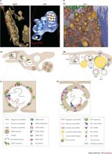
          • RNA replication of Flaviviridae family members occurs in tight association with endoplasmic reticulum-derived membranes, which are reorganized into viral replication organelles. Whereas the morphology and the architecture of these replication organelles are well defined, relatively little is known about the viral and cellular factors orchestrating their biogenesis.

          • Protein folding, modification and degradation are essential, tightly regulated cellular processes, and a number of common host factors and pathways that are involved in these processes appear to be exploited by both flaviviruses and hepaciviruses at different steps of their replication cycle. These include heat shock protein 70 (HSP70) network components, the unfolded protein response, the ubiquitin-dependent proteasome system and autophagy.

          • Accumulating evidence indicates that lipids and lipid metabolism fulfil essential roles in the life cycle of Flaviviridae viruses. They alter the lipid composition of cellular membranes, serving as scaffold for the assembly of the viral replicase by changing their biophysical properties, such as curvature, permeability and fluidity.

          • The identification of host cell pathways and factors commonly used by members of the Flaviviridae family might help in the development of broad-spectrum antiviral drugs that target multiple members of this family and/or other virus families.

          • As exemplified by members of the Flaviviridae family, the use of host cell pathways does not follow conventional phylogeny but, rather, reveals unexpected commonalities with distantly related viruses, raising the question of evolutionary relationships between these viruses.

          Supplementary information

          The online version of this article (doi:10.1038/nrmicro.2017.170) contains supplementary material, which is available to authorized users.

          Abstract

          In this Review, Bartenschlager and colleagues discuss how Flaviviridaeviruses rewire cellular pathways and co-opt organelles. They compare strategies employed by flaviviruses with those employed by hepaciviruses and discuss the importance of these virus–host interactions in the context of viral replication and antiviral therapies.

          Supplementary information

          The online version of this article (doi:10.1038/nrmicro.2017.170) contains supplementary material, which is available to authorized users.

          Abstract

          Members of the Flaviviridae virus family comprise a large group of enveloped viruses with a single-strand RNA genome of positive polarity. Several genera belong to this family, including the Hepacivirus genus, of which hepatitis C virus (HCV) is the prototype member, and the Flavivirus genus, which contains both dengue virus and Zika virus. Viruses of these genera differ in many respects, such as the mode of transmission or the course of infection, which is either predominantly persistent in the case of HCV or acutely self-limiting in the case of flaviviruses. Although the fundamental replication strategy of Flaviviridae members is similar, during the past few years, important differences have been discovered, including the way in which these viruses exploit cellular resources to facilitate viral propagation. These differences might be responsible, at least in part, for the various biological properties of these viruses, thus offering the possibility to learn from comparisons. In this Review, we discuss the current understanding of how Flaviviridae viruses manipulate and usurp cellular pathways in infected cells. Specifically, we focus on comparing strategies employed by flaviviruses with those employed by hepaciviruses, and we discuss the importance of these interactions in the context of viral replication and antiviral therapies.

          Supplementary information

          The online version of this article (doi:10.1038/nrmicro.2017.170) contains supplementary material, which is available to authorized users.

          Related collections

          Most cited references136

          • Record: found
          • Abstract: found
          • Article: not found

          Autophagy is activated for cell survival after endoplasmic reticulum stress.

          Eukaryotic cells deal with accumulation of unfolded proteins in the endoplasmic reticulum (ER) by the unfolded protein response, involving the induction of molecular chaperones, translational attenuation, and ER-associated degradation, to prevent cell death. Here, we found that the autophagy system is activated as a novel signaling pathway in response to ER stress. Treatment of SK-N-SH neuroblastoma cells with ER stressors markedly induced the formation of autophagosomes, which were recognized at the ultrastructural level. The formation of green fluorescent protein (GFP)-LC3-labeled structures (GFP-LC3 "dots"), representing autophagosomes, was extensively induced in cells exposed to ER stress with conversion from LC3-I to LC3-II. In IRE1-deficient cells or cells treated with c-Jun N-terminal kinase (JNK) inhibitor, the autophagy induced by ER stress was inhibited, indicating that the IRE1-JNK pathway is required for autophagy activation after ER stress. In contrast, PERK-deficient cells and ATF6 knockdown cells showed that autophagy was induced after ER stress in a manner similar to the wild-type cells. Disturbance of autophagy rendered cells vulnerable to ER stress, suggesting that autophagy plays important roles in cell survival after ER stress.
            Bookmark
            • Record: found
            • Abstract: found
            • Article: not found

            Composition and Three-Dimensional Architecture of the Dengue Virus Replication and Assembly Sites

            Summary Positive-strand RNA viruses are known to rearrange cellular membranes to facilitate viral genome replication. The biogenesis and three-dimensional organization of these membranes and the link between replication and virus assembly sites is not fully clear. Using electron microscopy, we find Dengue virus (DENV)-induced vesicles, convoluted membranes, and virus particles to be endoplasmic reticulum (ER)-derived, and we detect double-stranded RNA, a presumed marker of RNA replication, inside virus-induced vesicles. Electron tomography (ET) shows DENV-induced membrane structures to be part of one ER-derived network. Furthermore, ET reveals vesicle pores that could enable release of newly synthesized viral RNA and reveals budding of DENV particles on ER membranes directly apposed to vesicle pores. Thus, DENV modifies ER membrane structure to promote replication and efficient encapsidation of the genome into progeny virus. This architecture of DENV replication and assembly sites could explain the coordination of distinct steps of the flavivirus replication cycle.
              Bookmark
              • Record: found
              • Abstract: found
              • Article: not found

              Efficacy and Long-Term Safety of a Dengue Vaccine in Regions of Endemic Disease.

              A candidate tetravalent dengue vaccine is being assessed in three clinical trials involving more than 35,000 children between the ages of 2 and 16 years in Asian-Pacific and Latin American countries. We report the results of long-term follow-up interim analyses and integrated efficacy analyses.
                Bookmark

                Author and article information

                Contributors
                ralf.bartenschlager@med.uni-heidelberg.de
                Journal
                Nat Rev Microbiol
                Nat. Rev. Microbiol
                Nature Reviews. Microbiology
                Nature Publishing Group UK (London )
                1740-1526
                1740-1534
                12 February 2018
                2018
                : 16
                : 3
                : 125-142
                Affiliations
                [1 ]GRID grid.7700.0, ISNI 0000 0001 2190 4373, Department of Infectious Diseases, , Molecular Virology, Heidelberg University, ; Heidelberg, 69120 Germany
                [2 ]GRID grid.452463.2, German Center for Infection Research, Heidelberg Partner Site, ; Heidelberg, 69120 Germany
                Article
                BFnrmicro2017170
                10.1038/nrmicro.2017.170
                7097628
                29430005
                bfea540a-fcae-4f55-914f-b8044a3b6935
                © Nature Publishing Group, a division of Macmillan Publishers Limited. All Rights Reserved. 2018

                This article is made available via the PMC Open Access Subset for unrestricted research re-use and secondary analysis in any form or by any means with acknowledgement of the original source. These permissions are granted for the duration of the World Health Organization (WHO) declaration of COVID-19 as a global pandemic.

                History
                Categories
                Article
                Custom metadata
                © Springer Nature Limited 2018

                hepatitis c virus,viral pathogenesis,antivirals,west nile virus,virus-host interactions,dengue virus,pathogens,viral proteins

                Comments

                Comment on this article