10
views
0
recommends
+1 Recommend
0 collections
    0
    shares
      • Record: found
      • Abstract: found
      • Article: found
      Is Open Access

      Influence of corneal spherical aberration on prediction error of the Haigis-L formula

      research-article

      Read this article at

      Bookmark
          There is no author summary for this article yet. Authors can add summaries to their articles on ScienceOpen to make them more accessible to a non-specialist audience.

          Abstract

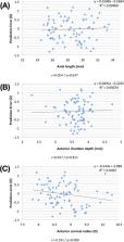
          The purpose of this study is to investigate the relationships between corneal asphericity and Haigis-L formula prediction errors in routine cataract surgery after refractive surgery for myopic correction. This retrospective study included 102 patients (102 eyes) with a history of previous PRK or LASIK and cataract surgery. Axial length, anterior chamber depth, and central corneal power were measured using the optical biometer. On the anterior corneal surface, Q-value, spherical aberration, and ecentricity at 6.0 and 8.0 mm were measured using a rotating Scheimpflug camera. The postoperative refractive outcome at 6 months, mean error, and mean absolute error were determined. Correlation tests were performed to determine the associations between pre-cataract surgery data and the prediction error. The Q-values for 6.0 and 8.0 mm corneal diameter were 1.57 ± 0.70 (range: 0.03~3.44), and 0.82 ± 0.5 (range: −0.10~−2.66). The spherical aberration for 6.0 and 8.0 mm diameter was 1.16 ± 0.39 µm (range: 0.24~2.08 µm), and 3.69 ± 0.87 µm (range: 0.91~5.91 µm). eccentricity for 6.0 and 8.0 mm diameter was −1.22 ± 0.31 (range: −1.85 to −0.17), and −0.82 ± 0.39 (range: −1.63 to 0.32). The spherical aberration for 8.0 mm cornea diameter showed the highest correlations with the predicion error (r = 0.750; p < 0.001). When the modified Haigis-L formula considering spherical aberration for 8.0 mm produced smaller values in standard deviation of mean error (0.45D versus 0.68D), mean absolute error (0.35D versus 0.55D), and median absolute error (0.31D versus 0.51D) than the Haigis formula. Corneal asphericity influences the predictive accuracy of the Haigis-L formula. The accuracy was enhanced by taking into consideration the corneal spherical aberration for the 8.0 mm zone at pre-cataract surgery state.

          Related collections

          Most cited references28

          • Record: found
          • Abstract: found
          • Article: not found

          Comparison of immersion ultrasound biometry and partial coherence interferometry for intraocular lens calculation according to Haigis.

          The precision of intraocular lens (IOL) calculation is essentially determined by the accuracy of the measurement of axial length. In addition to classical ultrasound biometry, partial coherence interferometry serves as a new optical method for axial length determination. A functional prototype from Carl Zeiss Jena implementing this principle was compared with immersion ultrasound biometry in our laboratory. In 108 patients attending the biometry laboratory for planning of cataract surgery, axial lengths were additionally measured optically. Whereas surgical decisions were based on ultrasound data, we used postoperative refraction measurements to calculate retrospectively what results would have been obtained if optical axial length data had been used for IOL calculation. For the translation of optical to geometrical lengths, five different conversion formulas were used, among them the relation which is built into the Zeiss IOL-Master. IOL calculation was carried out according to Haigis with and without optimization of constants. On the basis of ultrasound immersion data from our Grieshaber Biometric System (GBS), postoperative refraction after implantation of a Rayner IOL type 755 U was predicted correctly within +/- 1 D in 85.7% and within +/- 2 D in 99% of all cases. An analogous result was achieved with optical axial length data after suitable transformation of optical path lengths into geometrical distances. Partial coherence interferometry is a noncontact, user- and patient-friendly method for axial length determination and IOL planning with an accuracy comparable to that of high-precision immersion ultrasound.
            Bookmark
            • Record: found
            • Abstract: found
            • Article: not found

            Benchmark standards for refractive outcomes after NHS cataract surgery.

            To establish benchmark standards for refractive outcome after cataract surgery in the National Health Service when implementing the 2004 biometry guidelines of the Royal College of Ophthalmologists and customising A constants. Three cycles of prospective data were collected throughout the cataract care pathway on all patients using an electronic medical record system (Medisoft Ophthalmology), between January 2003 and February 2006. The electronic medical record automatically recommends the formula to be used according to the College guidelines and allows A constants to be customised separately for either ultrasound or partial coherence interferometry methods of axial length measurement and for different intraocular lens models. Consultants and trainees performed routine phacoemulsification cataract surgery and new intraocular lens models were introduced during the cycles. Uncomplicated cases with 'in-the-bag fixation', achieving 6/12 Snellen acuity or better were included. Community ophthalmic opticians performed refraction at 4 weeks. The postoperative subjective refraction was within 1 D of the predicted value in 79.7% of the 952 cases in cycle 1, 83.4% of 2406 cases in cycle 2, and 87.0% of 1448 cases in cycle 3. On the basis of our data, using College formula, optimising A constants and partial coherence interferometry, a benchmark standard of 85% of patients achieving a final spherical equivalent within 1 D of the predicted figure and 55% of patients within 0.5 D should be adopted.
              Bookmark
              • Record: found
              • Abstract: found
              • Article: not found

              Intraocular lens calculation after refractive surgery for myopia: Haigis-L formula.

              To describe the Haigis-L formula for the calculation of intraocular lenses (IOLs) after refractive laser surgery for myopia based on current biometry and keratometry and present clinical results. University Eye Hospital, Wuerzburg, Germany, and various clinics and private practices. The basic concepts of the new algorithm were described and summarized. The Haigis formula was analyzed with respect to its usability for eyes after laser surgery for myopia and modified accordingly. Correction curves for IOLMaster keratometry were derived from previous studies. The new formula was checked using the postoperative results of 187 cataract procedures in which 32 IOL types were implanted by 57 surgeons. Input data were current IOLMaster biometry as follows: axial length (AL), anterior chamber depth (ACD), and keratometry (corneal radii) measurements. Before IOL surgery, the mean spherical equivalent was -7.60 diopters (D)+/-3.90 (SD) (range -20.00 to -1.25 D); the mean AL, 27.02+/-2.01 mm (range 23.09 to 35.32 mm); the mean ACD, 3.52 +/- 0.36 mm (range 2.43 to 4.39 mm); and the mean of the measured corneal radii, 8.70+/-0.60 mm (range 7.28 to 10.96 mm). The mean arithmetic refractive prediction error was -0.04+/-0.70 D (range -2.30 to +2.40 D) and the median absolute error, 0.37 D (range +0.01 to +2.40 D). The percentages of correct refraction predictions within +/-2.00, +/-1.00, and +/-0.50 D were 98.4%, 84.0%, and 61.0%, respectively. The new formula would produce promising results in eyes without refractive history. Its refractive predictability fulfills the current criteria for normal eyes.
                Bookmark

                Author and article information

                Contributors
                gyoon@ur.rochester.edu
                Journal
                Sci Rep
                Sci Rep
                Scientific Reports
                Nature Publishing Group UK (London )
                2045-2322
                15 April 2020
                15 April 2020
                2020
                : 10
                : 6445
                Affiliations
                [1 ]ISNI 0000 0004 0470 4224, GRID grid.411947.e, Department of Ophthalmology, , Yeouido St. Mary’s Hospital, College of Medicine, The Catholic University of Korea, ; Seoul, Korea
                [2 ]ISNI 0000 0004 0647 8718, GRID grid.416981.3, Department Ophthalmology, , Uijeongbu St. Mary Hospital, College of Medicine, The Catholic University of Korea, ; Uijeongbu, Korea
                [3 ]ISNI 0000 0004 0470 4224, GRID grid.411947.e, Department of Ophthalmology, , Seoul St. Mary’s Hospital, College of Medicine, The Catholic University of Korea, ; Seoul, Korea
                [4 ]ISNI 0000 0004 1936 9174, GRID grid.16416.34, Flaum Eye Institute, Center for Visual Science, The Institute of Optics, University of Rochester, ; Rochester, New York USA
                Author information
                http://orcid.org/0000-0002-2018-5693
                http://orcid.org/0000-0001-6825-8612
                Article
                63594
                10.1038/s41598-020-63594-4
                7160126
                32296098
                c26eaed3-bcde-4e39-a668-d508f6862048
                © The Author(s) 2020

                Open Access This article is licensed under a Creative Commons Attribution 4.0 International License, which permits use, sharing, adaptation, distribution and reproduction in any medium or format, as long as you give appropriate credit to the original author(s) and the source, provide a link to the Creative Commons license, and indicate if changes were made. The images or other third party material in this article are included in the article’s Creative Commons license, unless indicated otherwise in a credit line to the material. If material is not included in the article’s Creative Commons license and your intended use is not permitted by statutory regulation or exceeds the permitted use, you will need to obtain permission directly from the copyright holder. To view a copy of this license, visit http://creativecommons.org/licenses/by/4.0/.

                History
                : 27 September 2019
                : 27 March 2020
                Categories
                Article
                Custom metadata
                © The Author(s) 2020

                Uncategorized
                lens diseases,outcomes research
                Uncategorized
                lens diseases, outcomes research

                Comments

                Comment on this article