29
views
0
recommends
+1 Recommend
0 collections
    0
    shares
      • Record: found
      • Abstract: found
      • Article: found
      Is Open Access

      Neural Mechanisms Underlying Conscious and Unconscious Gaze-Triggered Attentional Orienting in Autism Spectrum Disorder

      research-article

      Read this article at

      Bookmark
          There is no author summary for this article yet. Authors can add summaries to their articles on ScienceOpen to make them more accessible to a non-specialist audience.

          Abstract

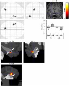
          Impaired joint attention represents the core clinical feature of autism spectrum disorder (ASD). Behavioral studies have suggested that gaze-triggered attentional orienting is intact in response to supraliminally presented eyes but impaired in response to subliminally presented eyes in individuals with ASD. However, the neural mechanisms underlying conscious and unconscious gaze-triggered attentional orienting remain unclear. We investigated this issue in ASD and typically developing (TD) individuals using event-related functional magnetic resonance imaging. The participants viewed cue stimuli of averted or straight eye gaze direction presented either supraliminally or subliminally and then localized a target. Reaction times were shorter when eye-gaze cues were directionally valid compared with when they were neutral under the supraliminal condition in both groups; the same pattern was found in the TD group but not the ASD group under the subliminal condition. The temporo–parieto–frontal regions showed stronger activation in response to averted eyes than to straight eyes in both groups under the supraliminal condition. The left amygdala was more activated while viewing averted vs. straight eyes in the TD group than in the ASD group under the subliminal condition. These findings provide an explanation for the neural mechanisms underlying the impairment in unconscious but not conscious gaze-triggered attentional orienting in individuals with ASD and suggest possible neurological and behavioral interventions to facilitate their joint attention behaviors.

          Related collections

          Most cited references71

          • Record: found
          • Abstract: found
          • Article: not found

          A multimodal cortical network for the detection of changes in the sensory environment.

          Sensory stimuli undergoing sudden changes draw attention and preferentially enter our awareness. We used event-related functional magnetic-resonance imaging (fMRI) to identify brain regions responsive to changes in visual, auditory and tactile stimuli. Unimodally responsive areas included visual, auditory and somatosensory association cortex. Multimodally responsive areas comprised a right-lateralized network including the temporoparietal junction, inferior frontal gyrus, insula and left cingulate and supplementary motor areas. These results reveal a distributed, multimodal network for involuntary attention to events in the sensory environment. This network contains areas thought to underlie the P300 event-related potential and closely corresponds to the set of cortical regions damaged in patients with hemineglect syndromes.
            Bookmark
            • Record: found
            • Abstract: found
            • Article: not found

            Social intelligence in the normal and autistic brain: an fMRI study.

            There is increasing support for the existence of 'social intelligence' [Humphrey (1984) Consciousness Regained], independent of general intelligence. Brothers et al. 1990) J. Cog. Neurosci., 4, 107-118] proposed a network of neural regions that comprise the 'social brain': the orbito-frontal cortex (OFC), superior temporal gyrus (STG) and amygdala. We tested Brothers' theory by examining both normal subjects as well as patients with high-functioning autism or Asperger syndrome (AS), who are well known to have deficits in social intelligence, and perhaps deficits in amygdala function [Bauman & Kemper (1988) J. Neuropath. Exp. Neurol., 47, 369]. We used a test of judging from the expressions of another person's eyes what that other person might be thinking or feeling. Using functional magnetic resonance imaging (fMRI) we confirmed Brothers' prediction that the STG and amygdala show increased activation when using social intelligence. Some areas of the prefrontal cortex also showed activation. In contrast, patients with autism or AS activated the fronto-temporal regions but not the amygdala when making mentalistic inferences from the eyes. These results provide support for the social brain theory of normal function, and the amygdala theory of autism.
              Bookmark
              • Record: found
              • Abstract: found
              • Article: not found

              Optimal experimental design for event-related fMRI.

              An important challenge in the design and analysis of event-related or single-trial functional magnetic resonance imaging (fMRI) experiments is to optimize statistical efficiency, i.e., the accuracy with which the event-related hemodynamic response to different stimuli can be estimated for a given amount of imaging time. Several studies have suggested that using a fixed inter-stimulus-interval (ISI) of at least 15 sec results in optimal statistical efficiency or power and that using shorter ISIs results in a severe loss of power. In contrast, recent studies have demonstrated the feasibility of using ISIs as short as 500 ms while still maintaining considerable efficiency or power. Here, we attempt to resolve this apparent contradiction by a quantitative analysis of the relative efficiency afforded by different event-related experimental designs. This analysis shows that statistical efficiency falls off dramatically as the ISI gets sufficiently short, if the ISI is kept fixed for all trials. However, if the ISI is properly jittered or randomized from trial to trial, the efficiency improves monotonically with decreasing mean ISI. Importantly, the efficiency afforded by such variable ISI designs can be more than 10 times greater than that which can be achieved by fixed ISI designs. These results further demonstrate the feasibility of using identical experimental designs with fMRI and electro-/magnetoencephalography (EEG/MEG) without sacrificing statistical power or efficiency of either technique, thereby facilitating comparison and integration across imaging modalities.
                Bookmark

                Author and article information

                Contributors
                Journal
                Front Hum Neurosci
                Front Hum Neurosci
                Front. Hum. Neurosci.
                Frontiers in Human Neuroscience
                Frontiers Media S.A.
                1662-5161
                28 June 2017
                2017
                : 11
                : 339
                Affiliations
                [1] 1Department of Neurodevelopmental Psychiatry, Habilitation and Rehabilitation, Graduate School of Medicine, Kyoto University Kyoto, Japan
                [2] 2Brain Activity Imaging Center, Advanced Telecommunications Research Institute International Kyoto, Japan
                [3] 3Faculty of Human Health Science, Graduate School of Medicine, Kyoto University Kyoto, Japan
                [4] 4The Organization for Promoting Neurodevelopmental Disorder Research Kyoto, Japan
                Author notes

                Edited by: Tal Kenet, Massachusetts General Hospital, United States

                Reviewed by: Karen Lisa Bales, University of California, Davis, United States; Elizabeth Redcay, University of Maryland, College Park, United States

                *Correspondence: Wataru Sato sato.wataru.4v@ 123456kyoto-u.ac.jp
                Article
                10.3389/fnhum.2017.00339
                5487428
                28701942
                c6128335-9c12-47cd-90a5-3f179a204237
                Copyright © 2017 Sato, Kochiyama, Uono, Yoshimura and Toichi.

                This is an open-access article distributed under the terms of the Creative Commons Attribution License (CC BY). The use, distribution or reproduction in other forums is permitted, provided the original author(s) or licensor are credited and that the original publication in this journal is cited, in accordance with accepted academic practice. No use, distribution or reproduction is permitted which does not comply with these terms.

                History
                : 20 March 2017
                : 12 June 2017
                Page count
                Figures: 4, Tables: 3, Equations: 0, References: 88, Pages: 13, Words: 9506
                Categories
                Neuroscience
                Original Research

                Neurosciences
                amygdala,attentional orienting,autism spectrum disorder (asd),eye gaze,functional magnetic resonance imaging (fmri),subliminal presentation

                Comments

                Comment on this article