72
views
0
recommends
+1 Recommend
0 collections
    0
    shares
      • Record: found
      • Abstract: found
      • Article: found
      Is Open Access

      The perils of global signal regression for group comparisons: a case study of Autism Spectrum Disorders

      research-article

      Read this article at

      Bookmark
          There is no author summary for this article yet. Authors can add summaries to their articles on ScienceOpen to make them more accessible to a non-specialist audience.

          Abstract

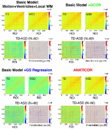
          We have previously argued from a theoretical basis that the standard practice of regression of the Global Signal from the fMRI time series in functional connectivity studies is ill advised, particularly when comparing groups of participants. Here, we demonstrate in resting-state data from participants with an Autism Spectrum Disorder and matched controls that these concerns are also well founded in real data. Using the prior theoretical work to formulate predictions, we show: (1) rather than simply altering the mean or range of correlation values amongst pairs of brain regions, Global Signal Regression systematically alters the rank ordering of values in addition to introducing negative values, (2) it leads to a reversal in the direction of group correlation differences relative to other preprocessing approaches, with a higher incidence of both long-range and local correlation differences that favor the Autism Spectrum Disorder group, (3) the strongest group differences under other preprocessing approaches are the ones most altered by Global Signal Regression, and (4) locations showing group differences no longer agree with those showing correlations with behavioral symptoms within the Autism Spectrum Disorder group. The correlation matrices of both participant groups under Global Signal Regression were well predicted by our previous mathematical analyses, demonstrating that there is nothing mysterious about these results. Finally, when independent physiological nuisance measures are lacking, we provide a simple alternative approach for assessing and lessening the influence of global correlations on group comparisons that replicates our previous findings. While this alternative performs less well for symptom correlations than our favored preprocessing approach that includes removal of independent physiological measures, it is preferable to the use of Global Signal Regression, which prevents unequivocal conclusions about the direction or location of group differences.

          Related collections

          Most cited references32

          • Record: found
          • Abstract: found
          • Article: not found

          An improved framework for confound regression and filtering for control of motion artifact in the preprocessing of resting-state functional connectivity data.

          Several recent reports in large, independent samples have demonstrated the influence of motion artifact on resting-state functional connectivity MRI (rsfc-MRI). Standard rsfc-MRI preprocessing typically includes regression of confounding signals and band-pass filtering. However, substantial heterogeneity exists in how these techniques are implemented across studies, and no prior study has examined the effect of differing approaches for the control of motion-induced artifacts. To better understand how in-scanner head motion affects rsfc-MRI data, we describe the spatial, temporal, and spectral characteristics of motion artifacts in a sample of 348 adolescents. Analyses utilize a novel approach for describing head motion on a voxelwise basis. Next, we systematically evaluate the efficacy of a range of confound regression and filtering techniques for the control of motion-induced artifacts. Results reveal that the effectiveness of preprocessing procedures on the control of motion is heterogeneous, and that improved preprocessing provides a substantial benefit beyond typical procedures. These results demonstrate that the effect of motion on rsfc-MRI can be substantially attenuated through improved preprocessing procedures, but not completely removed. Copyright © 2012 Elsevier Inc. All rights reserved.
            Bookmark
            • Record: found
            • Abstract: found
            • Article: not found

            Separating respiratory-variation-related fluctuations from neuronal-activity-related fluctuations in fMRI.

            Subtle changes in a subject's breathing rate or depth, which occur naturally during rest at low frequencies (<0.1 Hz), have been shown to be significantly correlated with fMRI signal changes throughout gray matter and near large vessels. The goal of this study was to investigate the impact of these low-frequency respiration variations on both task activation fMRI studies and resting-state functional connectivity analysis. Unlike MR signal changes correlated with the breathing motion ( approximately 0.3 Hz), BOLD signal changes correlated with across-breath variations in respiratory volume ( approximately 0.03 Hz) appear localized to blood vessels and regions with high blood volume, such as gray matter, similar to changes seen in response to a breath-hold challenge. In addition, the respiration-variation-induced signal changes were found to coincide with many of the areas identified as part of the 'default mode' network, a set of brain regions hypothesized to be more active at rest. Regions could therefore be classified as being part of a resting network based on their similar respiration-induced changes rather than their synchronized neuronal activity. Monitoring and removing these respiration variations led to a significant improvement in the identification of task-related activation and deactivation and only slight differences in regions correlated with the posterior cingulate at rest. Regressing out global signal changes or cueing the subject to breathe at a constant rate and depth resulted in an improved spatial overlap between deactivations and resting-state correlations among areas that showed deactivation.
              Bookmark
              • Record: found
              • Abstract: found
              • Article: not found

              Cortical activation and synchronization during sentence comprehension in high-functioning autism: evidence of underconnectivity.

              The brain activation of a group of high-functioning autistic participants was measured using functional MRI during sentence comprehension and the results compared with those of a Verbal IQ-matched control group. The groups differed in the distribution of activation in two of the key language areas. The autism group produced reliably more activation than the control group in Wernicke's (left laterosuperior temporal) area and reliably less activation than the control group in Broca's (left inferior frontal gyrus) area. Furthermore, the functional connectivity, i.e. the degree of synchronization or correlation of the time series of the activation, between the various participating cortical areas was consistently lower for the autistic than the control participants. These findings suggest that the neural basis of disordered language in autism entails a lower degree of information integration and synchronization across the large-scale cortical network for language processing. The article presents a theoretical account of the findings, related to neurobiological foundations of underconnectivity in autism.
                Bookmark

                Author and article information

                Journal
                Front Hum Neurosci
                Front Hum Neurosci
                Front. Hum. Neurosci.
                Frontiers in Human Neuroscience
                Frontiers Media S.A.
                1662-5161
                12 July 2013
                2013
                : 7
                : 356
                Affiliations
                [1] 1Section on Cognitive Neuropsychology, Laboratory of Brain and Cognition, National Institute of Mental Health, National Institutes of Health Bethesda, MD, USA
                [2] 2Scientific and Statistical Computing Core, National Institute of Mental Health, National Institutes of Health Bethesda, MD, USA
                Author notes

                Edited by: Ralph-Axel Müller, San Diego State University, USA

                Reviewed by: Michael Milham, Child Mind Institute, USA; Jeff Anderson, University of Utah, USA

                *Correspondence: Stephen J. Gotts, Laboratory of Brain and Cognition, National Institute of Mental Health, National Institutes of Health, Bldg. 10, Rm. 4C-217, Bethesda, MD 20892-1366, USA e-mail: gottss@ 123456mail.nih.gov
                Article
                10.3389/fnhum.2013.00356
                3709423
                23874279
                c8d2ee60-1313-4700-9724-21507466f751
                Copyright © 2013 Gotts, Saad, Jo, Wallace, Cox and Martin.

                This is an open-access article distributed under the terms of the Creative Commons Attribution License, which permits use, distribution and reproduction in other forums, provided the original authors and source are credited and subject to any copyright notices concerning any third-party graphics etc.

                History
                : 29 April 2013
                : 21 June 2013
                Page count
                Figures: 11, Tables: 0, Equations: 4, References: 68, Pages: 20, Words: 15760
                Categories
                Neuroscience
                Original Research Article

                Neurosciences
                functional connectivity,typically developing,artifact,resting-state fmri,gcor,global correlation

                Comments

                Comment on this article