5
views
0
recommends
+1 Recommend
0 collections
    0
    shares
      • Record: found
      • Abstract: found
      • Article: found
      Is Open Access

      Dysfunctional LAT2 Amino Acid Transporter Is Associated With Cataract in Mouse and Humans

      research-article
      1 , 2 , 3 , 4 , 5 , 1 , 2 , 5 , 6 , 7 , 8 , 4 , 6 , 9 , 7 , 1 , 2 , 10 , 11 , 12 , 13 , 4 , 5 , 14 , 12 , 2 , 7 , 15 , 16 , 12 , 17 , 18 , 10 , 11 , 4 , 5 , 14 , * , 5 , 6 , 19 , * , 1 , 2 , 3 , * , 7 , 8 , *
      Frontiers in Physiology
      Frontiers Media S.A.
      amino acid transporters LAT2 and TAT1, gene expression, cataract, ocular tissues, mouse model, patient screen

      Read this article at

      Bookmark
          There is no author summary for this article yet. Authors can add summaries to their articles on ScienceOpen to make them more accessible to a non-specialist audience.

          Abstract

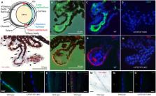
          Cataract, the loss of ocular lens transparency, accounts for ∼50% of worldwide blindness and has been associated with water and solute transport dysfunction across lens cellular barriers. We show that neutral amino acid antiporter LAT2 (Slc7a8) and uniporter TAT1 ( Slc16a10) are expressed on mouse ciliary epithelium and LAT2 also in lens epithelium. Correspondingly, deletion of LAT2 induced a dramatic decrease in lens essential amino acid levels that was modulated by TAT1 defect. Interestingly, the absence of LAT2 led to increased incidence of cataract in mice, in particular in older females, and a synergistic effect was observed with simultaneous lack of TAT1. Screening SLC7A8 in patients diagnosed with congenital or age-related cataract yielded one homozygous single nucleotide deletion segregating in a family with congenital cataract. Expressed in HeLa cells, this LAT2 mutation did not support amino acid uptake. Heterozygous LAT2 variants were also found in patients with cataract some of which showed a reduced transport function when expressed in HeLa cells. Whether heterozygous LAT2 variants may contribute to the pathology of cataract needs to be further investigated. Overall, our results suggest that defects of amino acid transporter LAT2 are implicated in cataract formation, a situation that may be aggravated by TAT1 defects.

          Related collections

          Most cited references46

          • Record: found
          • Abstract: found
          • Article: found
          Is Open Access

          Cat-Map: putting cataract on the map

          Lens opacities, or cataract(s), may be inherited as a classic Mendelian disorder usually with early-onset or, more commonly, acquired with age as a multi-factorial or complex trait. Many genetic forms of cataract have been described in mice and other animal models. Considerable progress has been made in mapping and identifying the genes and mutations responsible for inherited forms of cataract, and genetic determinants of age-related cataract are beginning to be discovered. To provide a convenient and accurate summary of current information focused on the increasing genetic complexity of Mendelian and age-related cataract we have created an online chromosome map and reference database for cataract in humans and mice (Cat-Map).
            Bookmark
            • Record: found
            • Abstract: found
            • Article: not found

            Identification of a membrane protein, LAT-2, that Co-expresses with 4F2 heavy chain, an L-type amino acid transport activity with broad specificity for small and large zwitterionic amino acids.

            We have identified a new human cDNA, L-amino acid transporter-2 (LAT-2), that induces a system L transport activity with 4F2hc (the heavy chain of the surface antigen 4F2, also named CD98) in oocytes. Human LAT-2 is the fourth member of the family of amino acid transporters that are subunits of 4F2hc. The amino acid transport activity induced by the co-expression of 4F2hc and LAT-2 was sodium-independent and showed broad specificity for small and large zwitterionic amino acids, as well as bulky analogs (e.g. BCH (2-aminobicyclo-(2,2,1)-heptane-2-carboxylic acid)). This transport activity was highly trans-stimulated, suggesting an exchanger mechanism of transport. Expression of tagged N-myc-LAT-2 alone in oocytes did not induce amino acid transport, and the protein had an intracellular location. Co-expression of N-myc-LAT-2 and 4F2hc gave amino acid transport induction and expression of N-myc-LAT-2 at the plasma membrane of the oocytes. These data suggest that LAT-2 is an additional member of the family of 4F2 light chain subunits, which associates with 4F2hc to express a system L transport activity with broad specificity for zwitterionic amino acids. Human LAT-2 mRNA is expressed in kidney > placenta > brain, liver > spleen, skeletal muscle, heart, small intestine, and lung. Human LAT-2 gene localizes at chromosome 14q11.2-13 (13 cR or approximately 286 kb from marker D14S1349). The high expression of LAT-2 mRNA in epithelial cells of proximal tubules, the basolateral location of 4F2hc in these cells, and the amino acid transport activity of LAT-2 suggest that this transporter contributes to the renal reabsorption of neutral amino acids in the basolateral domain of epithelial proximal tubule cells.
              Bookmark
              • Record: found
              • Abstract: found
              • Article: not found

              LAT2, a new basolateral 4F2hc/CD98-associated amino acid transporter of kidney and intestine.

              Glycoprotein-associated amino acid transporters (gpaAT) are permease-related proteins that require heterodimerization to express their function. So far, four vertebrate gpaATs have been shown to associate with 4F2hc/CD98 for functional expression, whereas one gpaAT specifically associates with rBAT. In this study, we characterized a novel gpaAT, LAT2, for which mouse and human cDNAs were identified by expressed sequence tag data base searches. The encoded ortholog proteins are 531 and 535 amino acids long and 92% identical. They share 52 and 48% residues with the gpaATs LAT1 and y(+)LAT1, respectively. When mouse LAT2 and human 4F2hc cRNAs were co-injected into Xenopus oocytes, disulfide-linked heterodimers were formed, and an L-type amino acid uptake was induced, which differed slightly from that produced by LAT1-4F2hc: the apparent affinity for L-phenylalanine was higher, and L-alanine was transported at physiological concentrations. In the presence of an external amino acid substrate, LAT2-4F2hc also mediated amino acid efflux. LAT2 mRNA is expressed mainly in kidney and intestine, whereas LAT1 mRNA is expressed widely. Immunofluorescence experiments showed colocalization of 4F2hc and LAT2 at the basolateral membrane of kidney proximal tubules and small intestine epithelia. In conclusion, LAT2 forms with LAT1 a subfamily of L-type gpaATs. We propose that LAT1 is involved in cellular amino acid uptake, whereas LAT2 plays a role in epithelial amino acid (re)absorption.
                Bookmark

                Author and article information

                Contributors
                Journal
                Front Physiol
                Front Physiol
                Front. Physiol.
                Frontiers in Physiology
                Frontiers Media S.A.
                1664-042X
                04 June 2019
                2019
                : 10
                : 688
                Affiliations
                [1] 1Institute of Physiology, University of Zurich , Zurich, Switzerland
                [2] 2Zurich Center for Integrative Human Physiology, University of Zurich , Zurich, Switzerland
                [3] 3Swiss National Centre of Competence in Research Kidney.CH, University of Zurich , Zurich, Switzerland
                [4] 4Genes, Disease and Therapy Program, Molecular Genetics Laboratory – IDIBELL , Barcelona, Spain
                [5] 5U730 and U731, Centro de Investigación Biomédica en Red de Enfermedades Raras , Barcelona, Spain
                [6] 6Institute for Research in Biomedicine, The Barcelona Institute of Science and Technology , Barcelona, Spain
                [7] 7Institute of Medical Molecular Genetics, University of Zurich , Zurich, Switzerland
                [8] 8Department of Biology, ETH Zurich , Zurich, Switzerland
                [9] 9Jules-Gonin Eye Hospital, University of Lausanne , Lausanne, Switzerland
                [10] 10Metabolomics Platform, IISPV, Department of Electronic Engineering, Universitat Rovira i Virgili , Tarragona, Spain
                [11] 11CIBER of Diabetes and Associated Metabolic Diseases (CIBERDEM) , Madrid, Spain
                [12] 12Andalusian Molecular Biology and Regenerative Medicine Centre – CABIMER , Seville, Spain
                [13] 13UCL Institute of Ophthalmology , London, United Kingdom
                [14] 14Genetics Section, Department of Physiological Sciences, Faculty of Medicine and Health Sciences, University of Barcelona , Barcelona, Spain
                [15] 15Neuroscience Center Zurich – ZNZ, University of Zurich and ETH Zurich , Zurich, Switzerland
                [16] 16Department of Ophthalmology and Vision Sciences, The Hospital for Sick Children , Toronto, ON, Canada
                [17] 17Moorfields Eye Hospital , London, United Kingdom
                [18] 18Department of Ophthalmology, School of Medicine, University of California, San Francisco , San Francisco, CA, United States
                [19] 19Departament de Bioquímica i Biomedicina Molecular, Facultat de Biologia, Universitat de Barcelona , Barcelona, Spain
                Author notes

                Edited by: Christine Kranz, University of Ulm, Germany

                Reviewed by: Snezana Petrovic, Wake Forest School of Medicine, United States; Yoshinori Marunaka, Kyoto Industrial Health Association, Japan

                *Correspondence: Virginia Nunes, vnunes@ 123456idibell.cat
                Barbara Kloeckener-Gruissem, barbarkl@ 123456ethz.ch

                These authors have contributed equally to this work

                These authors shared senior authorship

                This article was submitted to Renal and Epithelial Physiology, a section of the journal Frontiers in Physiology

                Article
                10.3389/fphys.2019.00688
                6558864
                31231240
                cdfdf93a-a59f-482c-ac49-e15b8ca5c54f
                Copyright © 2019 Knöpfel, Vilches, Camargo, Errasti-Murugarren, Stäubli, Mayayo, Munier, Miroshnikova, Poncet, Junza, Bhattacharya, Prat, Berry, Berger, Heon, Moore, Yanes, Nunes, Palacín, Verrey and Kloeckener-Gruissem.

                This is an open-access article distributed under the terms of the Creative Commons Attribution License (CC BY). The use, distribution or reproduction in other forums is permitted, provided the original author(s) and the copyright owner(s) are credited and that the original publication in this journal is cited, in accordance with accepted academic practice. No use, distribution or reproduction is permitted which does not comply with these terms.

                History
                : 27 March 2019
                : 16 May 2019
                Page count
                Figures: 5, Tables: 0, Equations: 0, References: 50, Pages: 12, Words: 0
                Categories
                Physiology
                Original Research

                Anatomy & Physiology
                amino acid transporters lat2 and tat1,gene expression,cataract,ocular tissues,mouse model,patient screen

                Comments

                Comment on this article