11
views
0
recommends
+1 Recommend
0 collections
    0
    shares
      • Record: found
      • Abstract: found
      • Article: found
      Is Open Access

      Potential Involvement of Platelet-Derived Microparticles and Microparticles Forming Immune Complexes during Monocyte Activation in Patients with Systemic Lupus Erythematosus

      research-article

      Read this article at

      Bookmark
          There is no author summary for this article yet. Authors can add summaries to their articles on ScienceOpen to make them more accessible to a non-specialist audience.

          Abstract

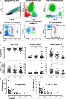
          Microparticles (MPs) are vesicles derived from the plasma membrane of different cells, are considered a source of circulating autoantigens, and can form immune complexes (MPs-ICs). The number of MPs and MPs-ICs increases in patients with systemic lupus erythematosus (SLE). MPs activate myeloid cells by inducing IL-6 and TNF-α in both SLE and other diseases. Therefore, we propose that the recognition of MPs-ICs by monocytes rather that MPs may define their phenotype and contribute to the inflammatory process in patients with SLE. Thus, the aims of this study were to evaluate the association among circulating MPs-ICs from different cell sources, alterations observed in monocyte subsets, and disease activity in patients with SLE and to establish whether monocytes bind and respond to MPs-ICs in vitro. Circulating MPs and monocyte subsets were characterized in 60 patients with SLE and 60 healthy controls (HCs) using multiparametric flow cytometry. Patients had higher MP counts and frequencies of MPs-CD41a + (platelet-derived) compared with HCs, regardless of disease activity. MPs from patients with SLE were C1q + and formed ICs with IgM and IgG. MPs-IgG + were positively correlated with active SLE (aSLE), whereas MPs-IgM + were negatively correlated. Most of the circulating total ICs-IgG + were located on MPs. The proportion and number of non-classical monocytes were significantly decreased in patients with SLE compared with HCs and in patients with aSLE compared with patients with the inactive disease. Non-classical monocytes obtained from patients with SLE exhibited increased levels of CD64 associated with MPs-IgG +, MPs-C1q +, total circulating ICs-IgG +, and disease activity. The direct effects of MPs and MPs-IgG + on monocytes were evaluated in cell culture. Monocytes from both HCs and patients bound to and internalized MPs and MPs-IgG + independent of CD64. These vesicles derived from platelets (PMPs), mainly PMPs-IgG +, activated monocytes in vitro and increased the expression of CD69, CD64, and pro-inflammatory cytokines such as IL-1β, TNF-α, and IFN-α. Therefore, MPs are one of the most representative sources of the total amount of circulating ICs-IgG + in patients with SLE. MPs-IgG + are associated with SLE activity, and PMPs-IgG + stimulate monocytes, changing their phenotype and promoting pro-inflammatory responses related to disease activity.

          Related collections

          Most cited references61

          • Record: found
          • Abstract: found
          • Article: not found

          Extracellular histones promote thrombin generation through platelet-dependent mechanisms: involvement of platelet TLR2 and TLR4.

          The release of histones from dying cells is associated with microvascular thrombosis and, because histones activate platelets, this could represent a possible pathogenic mechanism. In the present study, we assessed the influence of histones on the procoagulant potential of human platelets in platelet-rich plasma (PRP) and in purified systems. Histones dose-dependently enhanced thrombin generation in PRP in the absence of any trigger, as evaluated by calibrated automated thrombinography regardless of whether the contact phase was inhibited. Activation of coagulation required the presence of fully activatable platelets and was not ascribable to platelet tissue factor, whereas targeting polyphosphate with phosphatase reduced thrombin generation even when factor XII (FXII) was blocked or absent. In the presence of histones, purified polyphosphate was able to induce thrombin generation in plasma independently of FXII. In purified systems, histones induced platelet aggregation; P-selectin, phosphatidylserine, and FV/Va expression; and prothrombinase activity. Blocking platelet TLR2 and TLR4 with mAbs reduced the percentage of activated platelets and lowered the amount of thrombin generated in PRP. These data show that histone-activated platelets possess a procoagulant phenotype that drives plasma thrombin generation and suggest that TLR2 and TLR4 mediate the activation process.
            Bookmark
            • Record: found
            • Abstract: found
            • Article: not found

            Type I interferon in systemic lupus erythematosus and other autoimmune diseases.

            Different genetic alterations may lead to type I interferon (IFN) overproduction in human systemic lupus erythematosus (SLE). The increased bioavailability of type I IFN contributes to peripheral tolerance breakdown through the activation of immature myeloid dendritic cells (mDCs). IFN-matured mDCs activate autoreactive T cells. These cells, together with plasmacytoid DCs, help expand autoreactive B cells. IFN-matured DCs also activate cytotoxic CD8+ T cells, possibly increasing apoptotic cell availability. The capture of apoptotic cells by mDCs and of nucleic acid-containing immune complexes by plasmacytoid DCs and B cells amplifies the autoimmune reaction leading to disease manifestations. Genetic alterations in lineages other than B cells might explain other autoimmune syndromes where type I IFNs appear to be involved.
              Bookmark
              • Record: found
              • Abstract: found
              • Article: not found

              Platelets release mitochondria serving as substrate for bactericidal group IIA-secreted phospholipase A2 to promote inflammation.

              Mitochondrial DNA (mtDNA) is a highly potent inflammatory trigger and is reportedly found outside the cells in blood in various pathologies. Platelets are abundant in blood where they promote hemostasis. Although lacking a nucleus, platelets contain functional mitochondria. On activation, platelets produce extracellular vesicles known as microparticles. We hypothesized that activated platelets could also release their mitochondria. We show that activated platelets release respiratory-competent mitochondria, both within membrane-encapsulated microparticles and as free organelles. Extracellular mitochondria are found in platelet concentrates used for transfusion and are present at higher levels in those that induced acute reactions (febrile nonhemolytic reactions, skin manifestations, and cardiovascular events) in transfused patients. We establish that the mitochondrion is an endogenous substrate of secreted phospholipase A2 IIA (sPLA2-IIA), a phospholipase otherwise specific for bacteria, likely reflecting the ancestral proteobacteria origin of mitochondria. The hydrolysis of the mitochondrial membrane by sPLA2-IIA yields inflammatory mediators (ie, lysophospholipids, fatty acids, and mtDNA) that promote leukocyte activation. Two-photon microscopy in live transfused animals revealed that extracellular mitochondria interact with neutrophils in vivo, triggering neutrophil adhesion to the endothelial wall. Our findings identify extracellular mitochondria, produced by platelets, at the midpoint of a potent mechanism leading to inflammatory responses. © 2014 by The American Society of Hematology.
                Bookmark

                Author and article information

                Contributors
                Journal
                Front Immunol
                Front Immunol
                Front. Immunol.
                Frontiers in Immunology
                Frontiers Media S.A.
                1664-3224
                01 March 2018
                2018
                : 9
                : 322
                Affiliations
                [1] 1Grupo de Inmunología Celular e Inmunogenética, Instituto de Investigaciones Médicas, Facultad de Medicina, Universidad de Antioquia UdeA , Medellín, Colombia
                [2] 2Unidad de Citometría de Flujo, Sede de Investigación Universitaria, Universidad de Antioquia UdeA , Medellín, Colombia
                [3] 3Sección de Reumatología, Hospital Universitario San Vicente Fundación , Medellín, Colombia
                Author notes

                Edited by: Philippe Saas, INSERM UMR1098 Interactions Hôte-Greffon-Tumeur & Ingénierie Cellulaire et Génique, France

                Reviewed by: Johan Van Der Vlag, Radboud University Nijmegen, Netherlands; Adam Bruno Ceroi, Ghent University, Belgium

                *Correspondence: Diana Castaño, diana.castano@ 123456udea.edu.co

                Specialty section: This article was submitted to Inflammation, a section of the journal Frontiers in Immunology

                Article
                10.3389/fimmu.2018.00322
                5837989
                29545790
                ce10d48c-456d-472e-a515-4fda66571406
                Copyright © 2018 Burbano, Villar-Vesga, Orejuela, Muñoz, Vanegas, Vásquez, Rojas and Castaño.

                This is an open-access article distributed under the terms of the Creative Commons Attribution License (CC BY). The use, distribution or reproduction in other forums is permitted, provided the original author(s) and the copyright owner are credited and that the original publication in this journal is cited, in accordance with accepted academic practice. No use, distribution or reproduction is permitted which does not comply with these terms.

                History
                : 11 September 2017
                : 05 February 2018
                Page count
                Figures: 6, Tables: 1, Equations: 0, References: 81, Pages: 18, Words: 11718
                Funding
                Funded by: Departamento Administrativo de Ciencia, Tecnología e Innovación 10.13039/100007637
                Award ID: call 617-2013 (C. Burbano is recipient of a doctoral scholarship), 111565740575
                Funded by: Universidad de Antioquia 10.13039/501100005278
                Award ID: Sostenibilidad, Sistema Universitario de Investigaciones, CODI (2013-05-42869836)
                Categories
                Immunology
                Original Research

                Immunology
                microparticles,monocyte activation,immune complexes,systemic lupus erythematosus,monocyte subsets

                Comments

                Comment on this article