19
views
0
recommends
+1 Recommend
0 collections
    0
    shares
      • Record: found
      • Abstract: found
      • Article: found
      Is Open Access

      Myristoylation: An Important Protein Modification in the Immune Response

      review-article

      Read this article at

      Bookmark
          There is no author summary for this article yet. Authors can add summaries to their articles on ScienceOpen to make them more accessible to a non-specialist audience.

          Abstract

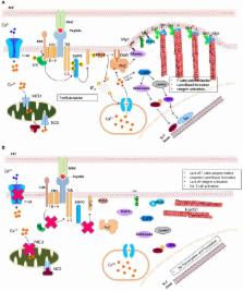
          Protein N-myristoylation is a cotranslational lipidic modification specific to the alpha-amino group of an N-terminal glycine residue of many eukaryotic and viral proteins. The ubiquitous eukaryotic enzyme, N-myristoyltransferase, catalyzes the myristoylation process. Precisely, attachment of a myristoyl group increases specific protein–protein interactions leading to subcellular localization of myristoylated proteins with its signaling partners. The birth of the field of myristoylation, a little over three decades ago, has led to the understanding of the significance of protein myristoylation in regulating cellular signaling pathways in several biological processes especially in carcinogenesis and more recently immune function. This review discusses myristoylation as a prerequisite step in initiating many immune cell signaling cascades. In particular, we discuss the hitherto unappreciated implication of myristoylation during myelopoiesis, innate immune response, lymphopoiesis for T cells, and the formation of the immunological synapse. Furthermore, we discuss the role of myristoylation in inducing the virological synapse during human immunodeficiency virus infection as well as its clinical implication. This review aims to summarize existing knowledge in the field and to highlight gaps in our understanding of the role of myristoylation in immune function so as to further investigate into the dynamics of myristoylation-dependent immune regulation.

          Related collections

          Most cited references145

          • Record: found
          • Abstract: found
          • Article: not found

          The interaction of Arp2/3 complex with actin: nucleation, high affinity pointed end capping, and formation of branching networks of filaments.

          The Arp2/3 complex is a stable assembly of seven protein subunits including two actin-related proteins (Arp2 and Arp3) and five novel proteins. Previous work showed that this complex binds to the sides of actin filaments and is concentrated at the leading edges of motile cells. Here, we show that Arp2/3 complex purified from Acanthamoeba caps the pointed ends of actin filaments with high affinity. Arp2/3 complex inhibits both monomer addition and dissociation at the pointed ends of actin filaments with apparent nanomolar affinity and increases the critical concentration for polymerization at the pointed end from 0.6 to 1.0 microM. The high affinity of Arp2/3 complex for pointed ends and its abundance in amoebae suggest that in vivo all actin filament pointed ends are capped by Arp2/3 complex. Arp2/3 complex also nucleates formation of actin filaments that elongate only from their barbed ends. From kinetic analysis, the nucleation mechanism appears to involve stabilization of polymerization intermediates (probably actin dimers). In electron micrographs of quick-frozen, deep-etched samples, we see Arp2/3 bound to sides and pointed ends of actin filaments and examples of Arp2/3 complex attaching pointed ends of filaments to sides of other filaments. In these cases, the angle of attachment is a remarkably constant 70 +/- 7 degrees. From these in vitro biochemical properties, we propose a model for how Arp2/3 complex controls the assembly of a branching network of actin filaments at the leading edge of motile cells.
            Bookmark
            • Record: found
            • Abstract: found
            • Article: not found

            Mechanism and function of formins in the control of actin assembly.

            Formins are a widely expressed family of proteins that govern cell shape, adhesion, cytokinesis, and morphogenesis by remodeling the actin and microtubule cytoskeletons. These large multidomain proteins associate with a variety of other cellular factors and directly nucleate actin polymerization through a novel mechanism. The signature formin homology 2 (FH2) domain initiates filament assembly and remains persistently associated with the fast-growing barbed end, enabling rapid insertion of actin subunits while protecting the end from capping proteins. On the basis of structural and mechanistic work, an integrated model is presented for FH2 processive motion. The adjacent FH1 domain recruits profilin-actin complexes and accelerates filament elongation. The most predominantly expressed formins in animals and fungi are autoinhibited through intramolecular interactions and appear to be activated by Rho GTPases and additional factors. Other classes of formins lack the autoinhibitory and/or Rho-binding domains and thus are likely to be controlled by alternative mechanisms.
              Bookmark
              • Record: found
              • Abstract: found
              • Article: not found

              Fatty acylation of proteins: new insights into membrane targeting of myristoylated and palmitoylated proteins.

              M Resh (1999)
              Covalent attachment of myristate and/or palmitate occurs on a wide variety of viral and cellular proteins. This review will highlight the latest advances in our understanding of the enzymology of N-myristoylation and palmitoylation as well as the functional consequences of fatty acylation of key signaling proteins. The role of myristate and palmitate in promoting membrane binding as well as specific membrane targeting will be reviewed, with emphasis on the Src family of tyrosine protein kinases and alpha subunits of heterotrimeric G proteins. The use of myristoyl switches and regulated depalmitoylation as mechanisms for achieving reversible membrane binding and regulated signaling will also be explored.
                Bookmark

                Author and article information

                Contributors
                URI : http://frontiersin.org/people/u/427886
                URI : http://frontiersin.org/people/u/435273
                URI : http://frontiersin.org/people/u/445707
                URI : http://frontiersin.org/people/u/453064
                URI : http://frontiersin.org/people/u/407593
                Journal
                Front Immunol
                Front Immunol
                Front. Immunol.
                Frontiers in Immunology
                Frontiers Media S.A.
                1664-3224
                30 June 2017
                2017
                : 8
                : 751
                Affiliations
                [1] 1Department of Biology, University of Winnipeg , Winnipeg, MB, Canada
                [2] 2Department of Biochemistry, University of Nigeria , Nsukka, Enugu, Nigeria
                [3] 3JC Wilt Infectious Diseases Research Institute, National HIV and Retrovirology Laboratory, Public Health Agency of Canada , Winnipeg, MB, Canada
                [4] 4Department of Medical Microbiology and Infectious Diseases, University of Manitoba , Winnipeg, MB, Canada
                [5] 5VastCon Inc. , Winnipeg, MB, Canada
                [6] 6Department of Biochemistry and Medical Genetics, University of Manitoba , Winnipeg, MB, Canada
                Author notes

                Edited by: Uday Kishore, Brunel University London, United Kingdom

                Reviewed by: Nadine Varin-Blank, Institut national de la santé et de la recherche médicale (INSERM), France; Zoltan Prohaszka, Semmelweis University, Hungary

                *Correspondence: Anuraag Shrivastav, a.shrivastav@ 123456uwinnipeg.ca

                Specialty section: This article was submitted to Molecular Innate Immunity, a section of the journal Frontiers in Immunology

                Article
                10.3389/fimmu.2017.00751
                5492501
                28713376
                d0b3666f-b966-4210-8fa7-4b09ae8fb15a
                Copyright © 2017 Udenwobele, Su, Good, Ball, Varma Shrivastav and Shrivastav.

                This is an open-access article distributed under the terms of the Creative Commons Attribution License (CC BY). The use, distribution or reproduction in other forums is permitted, provided the original author(s) or licensor are credited and that the original publication in this journal is cited, in accordance with accepted academic practice. No use, distribution or reproduction is permitted which does not comply with these terms.

                History
                : 27 January 2017
                : 13 June 2017
                Page count
                Figures: 5, Tables: 0, Equations: 0, References: 160, Pages: 16, Words: 11994
                Categories
                Immunology
                Review

                Immunology
                myristoylation,n-myristoyltransferase,lipid modification,signal transduction,t cells,human immunodeficiency virus

                Comments

                Comment on this article