36
views
0
recommends
+1 Recommend
0 collections
    0
    shares
      • Record: found
      • Abstract: found
      • Article: found
      Is Open Access

      Cartilage-Specific Overexpression of ERRγ Results in Chondrodysplasia and Reduced Chondrocyte Proliferation

      research-article

      Read this article at

      Bookmark
          There is no author summary for this article yet. Authors can add summaries to their articles on ScienceOpen to make them more accessible to a non-specialist audience.

          Abstract

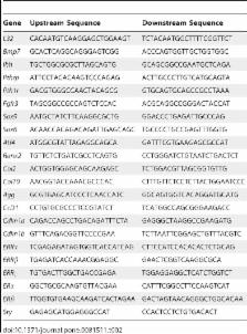
          While the role of estrogen receptor-related receptor alpha (ERRα) in chondrogenesis has been investigated, the involvement of ERR gamma (ERRγ) has not been determined. To assess the effect of increased ERRγ activity on cartilage development in vivo, we generated two transgenic (Tg) lines overexpressing ERRγ2 via a chondrocyte-specific promoter; the two lines exhibited ∼3 and ∼5 fold increased ERRγ2 protein expression respectively in E14.5 Tg versus wild type (WT) limbs. On postnatal day seven (P7), we observed a 4–10% reduction in the size of the craniofacial, axial and appendicular skeletons in Tg versus WT mice. The reduction in bone length was already present at birth and did not appear to involve bones that are derived via intramembranous bone formation as the bones of the calvaria, clavicle, and the mandible developed normally. Histological analysis of P7 growth plates revealed a reduction in the length of the Tg versus WT growth plate, the majority of which was attributable to a reduced proliferative zone. The reduced proliferative zone paralleled a decrease in the number of Ki67-positive proliferating cells, with no significant change in apoptosis, and was accompanied by large cell-free swaths of cartilage matrix, which extended through multiple zones of the growth plate. Using a bioinformatics approach, we identified known chondrogenesis-associated genes with at least one predicted ERR binding site in their proximal promoters, as well as cell cycle regulators known to be regulated by ERRγ. Of the genes identified, Col2al, Agg, Pth1r, and Cdkn1b (p27) were significantly upregulated, suggesting that ERRγ2 negatively regulates chondrocyte proliferation and positively regulates matrix synthesis to coordinate growth plate height and organization.

          Related collections

          Most cited references37

          • Record: found
          • Abstract: found
          • Article: not found

          Runx2 and Runx3 are essential for chondrocyte maturation, and Runx2 regulates limb growth through induction of Indian hedgehog.

          The differentiation of mesenchymal cells into chondrocytes and chondrocyte proliferation and maturation are fundamental steps in skeletal development. Runx2 is essential for osteoblast differentiation and is involved in chondrocyte maturation. Although chondrocyte maturation is delayed in Runx2-deficient (Runx2(-/-)) mice, terminal differentiation of chondrocytes does occur, indicating that additional factors are involved in chondrocyte maturation. We investigated the involvement of Runx3 in chondrocyte differentiation by generating Runx2-and-Runx3-deficient (Runx2(-/-)3(-/-)) mice. We found that chondrocyte differentiation was inhibited depending on the dosages of Runx2 and Runx3, and Runx2(-/-)3(-/-) mice showed a complete absence of chondrocyte maturation. Further, the length of the limbs was reduced depending on the dosages of Runx2 and Runx3, due to reduced and disorganized chondrocyte proliferation and reduced cell size in the diaphyses. Runx2(-/-)3(-/-) mice did not express Ihh, which regulates chondrocyte proliferation and maturation. Adenoviral introduction of Runx2 in Runx2(-/-) chondrocyte cultures strongly induced Ihh expression. Moreover, Runx2 directly bound to the promoter region of the Ihh gene and strongly induced expression of the reporter gene driven by the Ihh promoter. These findings demonstrate that Runx2 and Runx3 are essential for chondrocyte maturation and that Runx2 regulates limb growth by organizing chondrocyte maturation and proliferation through the induction of Ihh expression.
            Bookmark
            • Record: found
            • Abstract: found
            • Article: not found

            Promoter traps in embryonic stem cells: a genetic screen to identify and mutate developmental genes in mice.

            A general strategy for selecting insertion mutations in mice has been devised. Constructs lacking a promoter and including a beta-galactosidase gene, or a reporter gene encoding a protein with both beta-galactosidase and neomycin phosphotransferase activity, were designed so that activation of the reporter gene depends on its insertion within an active transcription unit. Such insertion events create a mutation in the tagged gene and allow its expression to be followed by beta-galactosidase activity. Introduction of promoter trap constructs into embryonic stem (ES) cells by electroporation or retroviral infection has led to the derivation of transgenic lines that show a variety of beta-galactosidase expression patterns. Intercrossing of heterozygotes from 24 strains that express beta-galactosidase identified 9 strains in which homozygosity leads to an embryonic lethality. Because no overt phenotype was detected in the remaining strains, these results suggest that a substantial proportion of mammalian genes identified by this approach are not essential for development.
              Bookmark
              • Record: found
              • Abstract: found
              • Article: not found

              ERRgamma directs and maintains the transition to oxidative metabolism in the postnatal heart.

              At birth, the heart undergoes a critical metabolic switch from a predominant dependence on carbohydrates during fetal life to a greater dependence on postnatal oxidative metabolism. This remains the principle metabolic state throughout life, although pathologic conditions such as heart failure and cardiac hypertrophy reactivate components of the fetal genetic program to increase carbohydrate utilization. Disruption of the ERRgamma gene (Esrrg), which is expressed at high levels in the fetal and postnatal mouse heart, blocks this switch, resulting in lactatemia, electrocardiographic abnormalities, and death during the first week of life. Genomic ChIP-on-chip and expression analysis identifies ERRgamma as both a direct and an indirect regulator of a nuclear-encoded mitochondrial genetic network that coordinates the postnatal metabolic transition. These findings reveal an unexpected and essential molecular genetic component of the oxidative metabolic gene program in the heart and highlight ERRgamma in the study of cardiac hypertrophy and failure.
                Bookmark

                Author and article information

                Contributors
                Role: Editor
                Journal
                PLoS One
                PLoS ONE
                plos
                plosone
                PLoS ONE
                Public Library of Science (San Francisco, USA )
                1932-6203
                2013
                9 December 2013
                : 8
                : 12
                : e81511
                Affiliations
                [1 ]Department of Medical Biophysics, University of Toronto, Toronto, Ontario, Canada
                [2 ]Department of Molecular Genetics, University of Toronto, Toronto, Ontario, Canada
                [3 ]Regenerative Medicine Program, Sprott Centre for Stem Cell Research, Ottawa Hospital Research Institute, Ottawa, Canada
                University of Texas Southwestern Medical Center, United States of America
                Author notes

                Competing Interests: The authors have declared that no competing interests exist.

                Conceived and designed the experiments: MC RAZ JFB KPM T-CT KT JEA. Performed the experiments: MC RAZ JFB KPM T-CT. Analyzed the data: MC RAZ JFB KPM T-CT KT JEA. Contributed reagents/materials/analysis tools: MC RAZ JFB KPM T-CT KT JEA. Wrote the paper: MC RAZ JFB KPM. Data interpretation: MC RAZ JFB KPM T-CT KT JEA. Revising manuscript content and approving final version of manuscript: MC RAZ JFB KPM T-CT KT JEA. Responsible for the integrity of the data analysis: MC RAZ JB KPM T-CT KT JEA.

                Article
                PONE-D-13-24404
                10.1371/journal.pone.0081511
                3857204
                d342564d-c9ef-4ae9-8a67-7b6afbeb7e19
                Copyright @ 2013

                This is an open-access article distributed under the terms of the Creative Commons Attribution License, which permits unrestricted use, distribution, and reproduction in any medium, provided the original author and source are credited.

                History
                : 14 June 2013
                : 14 October 2013
                Page count
                Pages: 11
                Funding
                This work was supported by a Canadian Institutes of Health Research (CIHR) operating grant (FRN 88104, JEA) and a Doctoral Research Award (MC); a Canadian Arthritis Network operating grant (JEA) and postdoctoral fellowship (RAZ), and a CIHR/Osteoporosis Society fellowship (RAZ). The funders had no role in study design, data collection and analysis, decision to publish, or preparation of the manuscript.
                Categories
                Research Article

                Uncategorized
                Uncategorized

                Comments

                Comment on this article