30
views
0
recommends
+1 Recommend
0 collections
    0
    shares
      • Record: found
      • Abstract: found
      • Article: found
      Is Open Access

      Sp1 expression regulates lung tumor progression

      research-article

      Read this article at

      Bookmark
          There is no author summary for this article yet. Authors can add summaries to their articles on ScienceOpen to make them more accessible to a non-specialist audience.

          Abstract

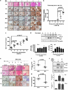
          The role of specificity protein 1 (Sp1) in controlling gene expression in lung tumor development and metastasis is not well understood. In this study, we showed that the Sp1 level was highly increased and required for lung tumor growth in transgenic mice bearing Kras-induced lung tumors under the control of doxycycline. Furthermore, the Sp1 level was highly upregulated in lung adenocarcinoma cells with low invasiveness and in patients with stage I lung cancer. We also demonstrated that Sp1 was downregulated in lung adenocarcinoma cells with high invasiveness and in patients with stage IV lung adenocarcinoma. Moreover, Sp1 inversely regulated migration, invasion and metastasis of lung adenocarcinoma cells in vivo. In addition, a decrease in the Sp1 level in highly invasive lung adenocarcinoma cells resulted from instability of the Sp1 protein. Furthermore, overexpression of Sp1 in highly invasive lung adenocarcinoma cells increased expression of E-cadherin, a suppressor of metastasis, and attenuated the translocation of β-catenin into the cellular nucleus that leads to tumor malignancy. Taken together, Sp1 level accumulated strongly in early stage and then declined in late stage, which is important for lung cancer cell proliferation and metastasis during tumorigenesis.

          Related collections

          Most cited references37

          • Record: found
          • Abstract: found
          • Article: not found

          Sp1 and krüppel-like factor family of transcription factors in cell growth regulation and cancer.

          The Sp/KLF family contains at least twenty identified members which include Sp1-4 and numerous krüppel-like factors. Members of the family bind with varying affinities to sequences designated as 'Sp1 sites' (e.g., GC-boxes, CACCC-boxes, and basic transcription elements). Family members have different transcriptional properties and can modulate each other's activity by a variety of mechanisms. Since cells can express multiple family members, Sp/KLF factors are likely to make up a transcriptional network through which gene expression can be fine-tuned. 'Sp1 site'-dependent transcription can be growth-regulated, and the activity, expression, and/or post-translational modification of multiple family members is altered with cell growth. Furthermore, Sp/KLF factors are involved in many growth-related signal transduction pathways and their overexpression can have positive or negative effects on proliferation. In addition to growth control, Sp/KLF factors have been implicated in apoptosis and angiogenesis; thus, the family is involved in several aspects of tumorigenesis. Consistent with a role in cancer, Sp/KLF factors interact with oncogenes and tumor suppressors, they can be oncogenic themselves, and altered expression of family members has been detected in tumors. Effects of changes in Sp/KLF factors are context-dependent and can appear contradictory. Since these factors act within a network, this diversity of effects may arise from differences in the expression profile of family members in various cells. Thus, it is likely that the properties of the overall network of Sp/KLF factors play a determining role in regulation of cell growth and tumor progression. Copyright 2001 Wiley-Liss, Inc.
            Bookmark
            • Record: found
            • Abstract: found
            • Article: not found

            Estrogen receptor analyses. Correlation of biochemical and immunohistochemical methods using monoclonal antireceptor antibodies.

            Attempts at histochemical localization of estrogen receptor with anti-steroid antibody or some fluoresceinated estrogens have given unacceptable sensitivities and specificities when compared with biochemical methods or clinical response. In the present study a monoclonal antibody against estrogen receptor (H222 Sp gamma) was used on cryostat sections of freshly frozen breast tumors with a peroxidase-antiperoxidase immunoperoxidase technique. Biochemical receptor analyses were by dextran-coated charcoal analyses. Tumors from three separate cohorts of patients were studied as follows: population A, 62 primary breast cancers from 1983; population B, 72 primary lesions stored from 1976 to 1983; and population C, 23 patients with metastases, treated with hormonal therapy. Distinct staining was seen in the cell nucleus. A semiquantitative relationship was seen between histochemical score assessment of staining and biochemical assay in each cohort. The sensitivity and specificity using a threshold of 75 for the histochemical score and more than 20 femtomoles/mg of protein for dextran-coated charcoal analyses were as follows: population A, specificity, 89%, and sensitivity, 95%; population B, specificity, 94%, and sensitivity 88%; and for population C, the comparison was with objective clinical response yielding specificity, 89%, and sensitivity, 93%.
              Bookmark
              • Record: found
              • Abstract: found
              • Article: not found

              Selection of invasive and metastatic subpopulations from a human lung adenocarcinoma cell line.

              To better understand the mechanism(s) underlying lung cancer invasion and metastasis, a Transwell invasion chamber was used to select progressively more invasive cancer cell populations from a clonal cell line of human lung adenocarcinoma, CL1. Five sublines with progressive invasiveness, designated CL1-1, CL1-2, CL1-3, CL1-4, and CL1-5, were obtained through this in vitro selection process. Their invasive abilities through basement membrane matrix showed a 4- to 6-fold increase over that of the parental cells. Moreover, the sublines manifested an increase in their colony-forming ability on soft agar, tumorigenicity, and metastatic potency in severe combined immunodeficiency (SCID) mice. Examining the phenotypes of the cell lines revealed increased expression of 92 kD gelatinase and an increase in the cell population stained with anti-keratin-8 and -18 antibodies. Clonal isolation of anti-keratin-18-antibody-positive and -negative cell populations demonstrated a correlated enhancement of the invasiveness of these cells and their expression of keratin-18. These results support the notion that the metastatic behavior of lung cancer cells can be characterized with this in vitro system, and that the properties of these progressively invasive cancer cells can be clonally studied.
                Bookmark

                Author and article information

                Journal
                Oncogene
                Oncogene
                Oncogene
                Nature Publishing Group
                0950-9232
                1476-5594
                30 August 2012
                12 December 2011
                : 31
                : 35
                : 3973-3988
                Affiliations
                [1 ]simpleInstitute of Basic Medical Sciences, College of Medicine, National Cheng-Kung University , Tainan, Taiwan
                [2 ]simpleDepartment of Pharmacology, College of Medicine, National Cheng-Kung University , Tainan, Taiwan
                [3 ]simpleInstitute of Bioinformatics and Biosignal Transduction, College of Bioscience in Biotechnology , simpleNational Cheng-Kung University , Tainan, Taiwan
                [4 ]simpleDepartment of Internal Medicine, College of Medicine and Hospital , simpleNational Cheng-Kung University , Tainan, Taiwan
                [5 ]simpleCenter for Gene Regulation and Signal Transduction, National Cheng-Kung University , Tainan, Taiwan
                [6 ]simpleGraduate Institute of Medical Sciences, College of Medicine, Taipei Medical University , Taipei, Taiwan
                Author notes
                [* ]simpleInstitute of Bioinformatics and Biosignal Transduction, College of Bioscience and Biotechnology, National Cheng-Kung University , No. 1, University Road, Tainan 701, Taiwan. E-mail: petehung@ 123456mail.ncku.edu.tw
                Article
                onc2011568
                10.1038/onc.2011.568
                3432230
                22158040
                d36e48b6-5c94-45e6-ad43-210817f756ad
                Copyright © 2012 Macmillan Publishers Limited

                This work is licensed under the Creative Commons Attribution-NonCommercial-No Derivative Works 3.0 Unported License. To view a copy of this license, visit http://creativecommons.org/licenses/by-nc-nd/3.0/

                History
                : 16 May 2011
                : 04 November 2011
                : 05 November 2011
                Categories
                Original Article

                Oncology & Radiotherapy
                sp1,metastasis,e-cadherin
                Oncology & Radiotherapy
                sp1, metastasis, e-cadherin

                Comments

                Comment on this article