8
views
0
recommends
+1 Recommend
0 collections
    0
    shares
      • Record: found
      • Abstract: found
      • Article: found
      Is Open Access

      A standardized and automated method of perineuronal net analysis using Wisteria floribunda agglutinin staining intensity

      research-article

      Read this article at

      Bookmark
          There is no author summary for this article yet. Authors can add summaries to their articles on ScienceOpen to make them more accessible to a non-specialist audience.

          Abstract

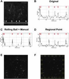
          Perineuronal nets (PNNs) are aggregations of extracellular matrix molecules that are critical for plasticity. Their altered development or changes during adulthood appear to contribute to a wide range of diseases/disorders of the brain. An increasing number of studies examining the contribution of PNN to various behaviors and types of plasticity have analyzed the fluorescence intensity of Wisteria floribunda agglutinin (WFA) as an indirect measure of the maturity of PNNs, with brighter WFA staining corresponding to a more mature PNN and dim WFA staining corresponding to an immature PNN. However, a clearly-defined and unified method for assessing the intensity of PNNs is critical to allow us to make comparisons across studies and to advance our understanding of how PNN plasticity contributes to normal brain function and brain disease states. Here we examined methods of PNN intensity quantification and demonstrate that creating a region of interest around each PNN and subtracting appropriate background is a viable method for PNN intensity quantification that can be automated. This method produces less variability and bias across experiments compared to other published analyses, and this method increases reproducibility and reliability of PNN intensity measures, which is critical for comparisons across studies in this emerging field.

          Highlights

          • PNN intensity quantification varies across the field.

          • The “ROI” method decreases variability among researchers.

          • The “ROI” method can be automated.

          Related collections

          Most cited references27

          • Record: found
          • Abstract: found
          • Article: not found

          Perineuronal nets protect fear memories from erasure.

          In adult animals, fear conditioning induces a permanent memory that is resilient to erasure by extinction. In contrast, during early postnatal development, extinction of conditioned fear leads to memory erasure, suggesting that fear memories are actively protected in adults. We show here that this protection is conferred by extracellular matrix chondroitin sulfate proteoglycans (CSPGs) in the amygdala. The organization of CSPGs into perineuronal nets (PNNs) coincided with the developmental switch in fear memory resilience. In adults, degradation of PNNs by chondroitinase ABC specifically rendered subsequently acquired fear memories susceptible to erasure. This result indicates that intact PNNs mediate the formation of erasure-resistant fear memories and identifies a molecular mechanism closing a postnatal critical period during which traumatic memories can be erased by extinction.
            Bookmark
            • Record: found
            • Abstract: not found
            • Article: not found

            Biomedical Image Processing

            Sternberg (1983)
              Bookmark
              • Record: found
              • Abstract: found
              • Article: not found

              The perineuronal net and the control of CNS plasticity.

              Perineuronal nets (PNNs) are reticular structures that surround the cell body of many neurones, and extend along their dendrites. They are considered to be a specialized extracellular matrix in the central nervous system (CNS). PNN formation is first detected relatively late in development, as the mature synaptic circuitry of the CNS is established and stabilized. Its unique distribution in different CNS regions, the timing of its establishment, and the changes it undergoes after injury all point toward diverse and important functions that it may be performing. The involvement of PNNs in neuronal plasticity has been extensively studied over recent years, with developmental, behavioural, and functional correlations. In this review, we will first briefly detail the structure and organization of PNNs, before focusing our discussion on their unique roles in neuronal development and plasticity. The PNN is an important regulator of CNS plasticity, both during development and into adulthood. Production of critical PNN components is often triggered by appropriate sensory experiences during early postnatal development. PNN deposition around neurones helps to stabilize the established neuronal connections, and to restrict the plastic changes due to future experiences within the CNS. Disruption of PNNs can reactivate plasticity in many CNSs, allowing activity-dependent changes to once again modify neuronal connections. The mechanisms through which PNNs restrict CNS plasticity remain unclear, although recent advances promise to shed additional light on this important subject.
                Bookmark

                Author and article information

                Contributors
                Journal
                IBRO Rep
                IBRO Rep
                IBRO Reports
                Elsevier
                2451-8301
                01 October 2016
                December 2016
                01 October 2016
                : 1
                : 54-60
                Affiliations
                [1]Department of Integrative Physiology and Neuroscience, Washington State University, Vancouver, WA 98686, USA
                Author notes
                []Corresponding author. Department of Integrative Physiology and Neuroscience, Translational Addiction Research Center, Washington State University, 14204 NE Salmon Creek Ave, Vancouver, WA 98686, USA. sorg@ 123456vetmed.wsu.edu
                Article
                S2451-8301(16)30013-9
                10.1016/j.ibror.2016.10.001
                5507617
                28713865
                d47cd919-79f9-42de-a232-d59b857e740a
                © 2016 The Authors

                This is an open access article under the CC BY-NC-ND license (http://creativecommons.org/licenses/by-nc-nd/4.0/).

                History
                : 5 September 2016
                : 1 October 2016
                : 1 October 2016
                Categories
                Article

                extracellular matrix,perineuronal net,wisteria floribunda agglutinin (wfa),automated image analysis,au, arbitrary unit,cpu, caudate/putamen,ecm, extracellular matrix,hip, dorsal hippocampus,nan, not a number,of, orbitofrontal cortex,pl, prelimbic,pfc, prefrontal cortex,pnn, perineuronal net,roi, region of interest,sd, standard deviation,sem, standard error of the mean,wfa, wisteria floribunda agglutinin

                Comments

                Comment on this article