8
views
0
recommends
+1 Recommend
0 collections
    0
    shares
      • Record: found
      • Abstract: found
      • Article: found
      Is Open Access

      Synapse-Selective Control of Cortical Maturation and Plasticity by Parvalbumin-Autonomous Action of SynCAM 1

      research-article
      1 , * , 2 , 1 , 3 , *
      Cell reports

      Read this article at

      Bookmark
          There is no author summary for this article yet. Authors can add summaries to their articles on ScienceOpen to make them more accessible to a non-specialist audience.

          SUMMARY

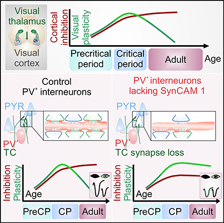
          Cortical plasticity peaks early in life and tapers in adulthood, as exemplified in the primary visual cortex (V1), wherein brief loss of vision in one eye reduces cortical responses to inputs from that eye during the critical period but not in adulthood. The synaptic locus of cortical plasticity and the cellautonomous synaptic factors determining critical periods remain unclear. We here demonstrate that the immunoglobulin protein Synaptic Cell Adhesion Molecule 1 (SynCAM 1/Cadm1) is regulated by visual experience and limits V1 plasticity. Loss of SynCAM 1 selectively reduces the number of thalamocortical inputs onto parvalbumin (PV +) interneurons, impairing the maturation of feedforward inhibition in V1. SynCAM 1 acts in PV + interneurons to actively restrict cortical plasticity, and brief PV +-specific knockdown of SynCAM 1 in adult visual cortex restores juvenile-like plasticity. These results identify a synapse-specific, cell-autonomous mechanism for thalamocortical visual circuit maturation and closure of the visual critical period.

          In Brief

          Ribic et al. show that cortical plasticity is actively restricted by the synapseorganizing molecule SynCAM 1. The protein acts in parvalbumin interneurons to recruit excitatory thalamocortical terminals. This controls the maturation of inhibition and actively limits cortical plasticity, revealing a synaptic locus for closure of cortical critical periods.

          Graphical Abstract

          Related collections

          Most cited references53

          • Record: found
          • Abstract: found
          • Article: not found

          Highly selective receptive fields in mouse visual cortex.

          Genetic methods available in mice are likely to be powerful tools in dissecting cortical circuits. However, the visual cortex, in which sensory coding has been most thoroughly studied in other species, has essentially been neglected in mice perhaps because of their poor spatial acuity and the lack of columnar organization such as orientation maps. We have now applied quantitative methods to characterize visual receptive fields in mouse primary visual cortex V1 by making extracellular recordings with silicon electrode arrays in anesthetized mice. We used current source density analysis to determine laminar location and spike waveforms to discriminate putative excitatory and inhibitory units. We find that, although the spatial scale of mouse receptive fields is up to one or two orders of magnitude larger, neurons show selectivity for stimulus parameters such as orientation and spatial frequency that is near to that found in other species. Furthermore, typical response properties such as linear versus nonlinear spatial summation (i.e., simple and complex cells) and contrast-invariant tuning are also present in mouse V1 and correlate with laminar position and cell type. Interestingly, we find that putative inhibitory neurons generally have less selective, and nonlinear, responses. This quantitative description of receptive field properties should facilitate the use of mouse visual cortex as a system to address longstanding questions of visual neuroscience and cortical processing.
            Bookmark
            • Record: found
            • Abstract: found
            • Article: not found

            Imaging large-scale neural activity with cellular resolution in awake, mobile mice.

            We report a technique for two-photon fluorescence imaging with cellular resolution in awake, behaving mice with minimal motion artifact. The apparatus combines an upright, table-mounted two-photon microscope with a spherical treadmill consisting of a large, air-supported Styrofoam ball. Mice, with implanted cranial windows, are head restrained under the objective while their limbs rest on the ball's upper surface. Following adaptation to head restraint, mice maneuver on the spherical treadmill as their heads remain motionless. Image sequences demonstrate that running-associated brain motion is limited to approximately 2-5 microm. In addition, motion is predominantly in the focal plane, with little out-of-plane motion, making the application of a custom-designed Hidden-Markov-Model-based motion correction algorithm useful for postprocessing. Behaviorally correlated calcium transients from large neuronal and astrocytic populations were routinely measured, with an estimated motion-induced false positive error rate of <5%.
              Bookmark
              • Record: found
              • Abstract: found
              • Article: not found

              The expression of vesicular glutamate transporters defines two classes of excitatory synapse.

              The quantal release of glutamate depends on its transport into synaptic vesicles. Recent work has shown that a protein previously implicated in the uptake of inorganic phosphate across the plasma membrane catalyzes glutamate uptake by synaptic vesicles. However, only a subset of glutamate neurons expresses this vesicular glutamate transporter (VGLUT1). We now report that excitatory neurons lacking VGLUT1 express a closely related protein that has also been implicated in phosphate transport. Like VGLUT1, this protein localizes to synaptic vesicles and functions as a vesicular glutamate transporter (VGLUT2). The complementary expression of VGLUT1 and 2 defines two distinct classes of excitatory synapse.
                Bookmark

                Author and article information

                Journal
                101573691
                39703
                Cell Rep
                Cell Rep
                Cell reports
                2211-1247
                11 January 2019
                08 January 2019
                24 January 2019
                : 26
                : 2
                : 381-393.e6
                Affiliations
                [1 ]Department of Neuroscience, Tufts University School of Medicine, Boston, MA 02111, USA
                [2 ]Department of Neuroscience, Yale University School of Medicine, New Haven, CT 06510, USA
                [3 ]Lead Contact
                Author notes

                AUTHOR CONTRIBUTIONS

                Conceptualization, A.R.; Methodology, A.R.; Software, A.R.; Investigation, A.R.; Writing – Original Draft, A.R.; Writing – Review & Editing, A.R. and T.B.; Funding Acquisition, A.R. and T.B.; Resources, M.C.C. and T.B.; Supervision, T.B.

                Article
                NIHMS1518268
                10.1016/j.celrep.2018.12.069
                6345548
                30625321
                befdad33-607e-43ef-a1a4-b8cecb7da156

                This is an open access article under the CC BY-NC-ND license ( http://creativecommons.org/licenses/by-nc-nd/4.0/).

                History
                Categories
                Article

                Cell biology
                Cell biology

                Comments

                Comment on this article