- Record: found
- Abstract: found
- Article: found
Is there a role of immunosenescence in the pathogenesis of malignant mesothelioma? A case study
letter
Read this article at
There is no author summary for this article yet. Authors can add summaries to their articles on ScienceOpen to make them more accessible to a non-specialist audience.
Abstract
Sir,
Malignant pleural mesothelioma is the most common cause of primary pleural malignancy.
Approximately, 35% of effusions associated with it are described to be inflammatory/reactive/lymphocytic
in nature.[1] The latency period, defined as the time between the first exposure to
asbestos and the development of mesothelioma, has been reported to be 40 years on
an average. However, latency periods as long as 72 years have been documented.[2]
Here, we present an interesting case of malignant mesothelioma in a 90-year-old female
with a remote and minimal history of exposure to asbestos. The case is quite interesting
because this is one of the longest latency periods ever reported.
A 90-year-old female with a history of bronchiectasis and chronic pseudomonas infection,
prior Mycobacterium avium intracellulare infection, pulmonary arterial hypertension,
and atrial fibrillation was seen in the clinic for increasing shortness of breath
over a period of 5 days. A chest X-ray revealed a large left-sided pleural effusion
that was considerably larger in size compared to 8 months back. A thoracentesis was
performed after admission which revealed yellow colored hazy fluid. A total of 1200
cc of pleural fluid was aspirated from the left pleural space under ultrasound guidance.
The fluid analysis revealed a lymphocyte predominant exudative fluid [Table 1]. The
differential diagnosis for the lymphocyte predominant fluid is narrow and includes
the following-Tuberculosis, sarcoidosis, lymphoma, yellow nail syndrome, chylothorax,
and rheumatoid pleurisy. Flow cytometry was performed which excluded lymphoma and
demonstrated a CD4 to CD8 ratio of 12:1. The clinical picture and result of the fluid
analysis excluded chylothorax, yellow nail syndrome, and rheumatoid arthritis as possible
causes. Further immune-histochemical evaluation of the pleural fluid revealed cells
that were positive for Calretinin and CD68, and negative for Ber-EP4, supporting a
reactive process. Malignant cells were not encountered. Post-procedure computed tomography
scans revealed a small hydropneumothorax, and to our surprise, multiple left-sided
pleural-based soft tissue masses [Figures 1 and 2]. A single chest wall implant was
also noted. A transthoracic needle biopsy from the mass was performed, followed by
a small-bore indwelling pleural catheter catheter placement. It demonstrated large
epithelioid tumor cells in cords, nests and tubular glandular structures [Figure 3].
These tumor cells were immunoreactive for cytokeratin AE1/AE3, calretinin, cytokeratin
5/6, WT1, D2-40 [Figure 4] and negative for thyroid transcription factor-1, and Napsin
A. This immunoreactive pattern was consistent with mesothelioma. A nonaggressive course
of treatment, focusing on comfort care was preferred by the patient. A more detailed
history, obtained after this rather unexpected diagnosis, revealed that the patient
was employed as a messenger at naval yard in 1940s about 74 years back. Any further
exposure to asbestos was ruled out. Based on this history of rather indirect exposure
to asbestos, and that too with a long latent period of >70 years, we felt that it
would be worthwhile to review the cause-and-effect relationship between asbestos and
mesothelioma.
Table 1
Results of pleural fluid analysis
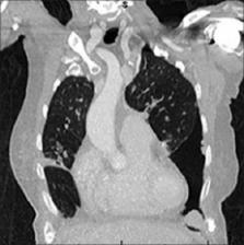
Figure 1
Coronal view demonstrating left pleural based multiple masses. A large left pneumothorax
is seen
Figure 2
Mediastinal window demonstrating a posterior lobulated mass along with pleural effusion
that tracks along the fissure. A small juxta-pericardial pleural based mass is also
noted on this view
Figure 3
This microscopic image (×200) shows sheets of epithelioid cells in an infiltrating
pattern typical of epithelioid mesothelioma
Figure 4
Immunohistochemistry is positive for WT 1 (a) (×200) and D2-40 (b) (×400) stains.
Greater than 90% of mesotheliomas stain positive for both these markers which are
negative in other lung cancers thus helping to distinguish between them
One explanation for such a long latency period could be that minimal exposure to asbestos
resulting in delayed development of cancer. It is also interesting to note that mesothelioma
can occur among immunocompromised people even without any history of exposure to asbestos.
We feel that age-related frailty of the immune system might explain the development
of mesothelioma in her case. Lending support to this hypothesis would be the absence
of pleural plaques/calcifications.
We reviewed some of the principal concepts of tumorigenesis associated with asbestos
exposure. Even though the available data is unclear, the intensity of exposure and
latency periods are commonly assumed to be inversely related in those who develop
cancer.[3
4] The risk of cancer development is related to the intensity of exposure. The duration
of exposure, even though considered to be less important, is also related to the risk
of cancer.[5] Thus, chronic low-level exposure can account for the development of
cancer. It is postulated that cancer will develop when the exposure to asbestos has
reached a certain degree, which varies between individuals.[3] It is also thought
that the failure of the body's immunological surveillance system to detect and kill
cancer cells results in the development of cancer.[6] The reports of mesothelioma
being related to HIV/AIDS, simian virus 40 infection, organ transplant, or advanced
age lends credence to the theory.[5]
Cancer is the result of the interplay of multiple factors: Exposure to asbestos as
well as the way the immune system responds to it. Considering the low-degree of exposure
and development of cancer after such a long period, this case provides support for
the role of immunosenescence in the development of mesothelioma.
Financial support and sponsorship
Nil.
Conflicts of interest
There are no conflicts of interest.
Related collections
Most cited references6
- Record: found
- Abstract: found
- Article: not found
Malignant mesothelioma: global incidence and relationship with asbestos.
Claudio Bianchi, Tommaso Bianchi (2007)
- Record: found
- Abstract: found
- Article: not found
Latency periods in asbestos-related mesothelioma of the pleura.
L Giarelli, A Brollo, C Zuch … (1997)
- Record: found
- Abstract: found
- Article: not found
A diagnosis of malignant pleural mesothelioma can be made by effusion cytology: results of a 20 year audit.
J. Olsen, Anna Nowak, K Shilkin … (2012)