10
views
0
recommends
+1 Recommend
0 collections
    0
    shares
      • Record: found
      • Abstract: found
      • Article: found
      Is Open Access

      Propofol inhibits prokaryotic voltage-gated Na + channels by promoting activation-coupled inactivation

      research-article

      Read this article at

      Bookmark
          There is no author summary for this article yet. Authors can add summaries to their articles on ScienceOpen to make them more accessible to a non-specialist audience.

          Abstract

          Despite extensive use in clinical practice, the mechanisms of propofol action on sodium channels are not fully understood. Yang et al. incorporate complementary biophysical approaches (electrophysiology and molecular dynamics simulations) to demonstrate that propofol inhibits two prokaryotic voltage-gated sodium channels, NaChBac and NavMs, by modulating both activation and inactivation gating.

          Abstract

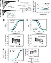
          Propofol is widely used in the clinic for the induction and maintenance of general anesthesia. As with most general anesthetics, however, our understanding of its mechanism of action remains incomplete. Local and general anesthetics largely inhibit voltage-gated Na + channels (Navs) by inducing an apparent stabilization of the inactivated state, associated in some instances with pore block. To determine the biophysical and molecular basis of propofol action in Navs, we investigated NaChBac and NavMs, two prokaryotic Navs with distinct voltage dependencies and gating kinetics, by whole-cell patch clamp electrophysiology in the absence and presence of propofol at clinically relevant concentrations (2–10 µM). In both Navs, propofol induced a hyperpolarizing shift of the pre-pulse inactivation curve without any significant effects on recovery from inactivation at strongly hyperpolarized voltages, demonstrating that propofol does not stabilize the inactivated state. Moreover, there was no evidence of fast or slow pore block by propofol in a non-inactivating NaChBac mutant (T220A). Propofol also induced hyperpolarizing shifts of the conductance-voltage relationships with negligible effects on the time constants of deactivation at hyperpolarized voltages, indicating that propofol does not stabilize the open state. Instead, propofol decreases the time constants of macroscopic activation and inactivation. Adopting a kinetic scheme of Nav gating that assumes preferential closed-state recovery from inactivation, a 1.7-fold acceleration of the rate constant of activation and a 1.4-fold acceleration of the rate constant of inactivation were sufficient to reproduce experimental observations with computer simulations. In addition, molecular dynamics simulations and molecular docking suggest that propofol binding involves interactions with gating machinery in the S4–S5 linker and external pore regions. Our findings show that propofol is primarily a positive gating modulator of prokaryotic Navs, which ultimately inhibits the channels by promoting activation-coupled inactivation.

          Related collections

          Most cited references81

          • Record: found
          • Abstract: found
          • Article: not found

          General anaesthesia: from molecular targets to neuronal pathways of sleep and arousal.

          The mechanisms through which general anaesthetics, an extremely diverse group of drugs, cause reversible loss of consciousness have been a long-standing mystery. Gradually, a relatively small number of important molecular targets have emerged, and how these drugs act at the molecular level is becoming clearer. Finding the link between these molecular studies and anaesthetic-induced loss of consciousness presents an enormous challenge, but comparisons with the features of natural sleep are helping us to understand how these drugs work and the neuronal pathways that they affect. Recent work suggests that the thalamus and the neuronal networks that regulate its activity are the key to understanding how anaesthetics cause loss of consciousness.
            Bookmark
            • Record: found
            • Abstract: found
            • Article: not found

            Voltage sensor of Kv1.2: structural basis of electromechanical coupling.

            Voltage-dependent ion channels contain voltage sensors that allow them to switch between nonconductive and conductive states over the narrow range of a few hundredths of a volt. We investigated the mechanism by which these channels sense cell membrane voltage by determining the x-ray crystal structure of a mammalian Shaker family potassium ion (K+) channel. The voltage-dependent K+ channel Kv1.2 grew three-dimensional crystals, with an internal arrangement that left the voltage sensors in an apparently native conformation, allowing us to reach three important conclusions. First, the voltage sensors are essentially independent domains inside the membrane. Second, they perform mechanical work on the pore through the S4-S5 linker helices, which are positioned to constrict or dilate the S6 inner helices of the pore. Third, in the open conformation, two of the four conserved Arg residues on S4 are on a lipid-facing surface and two are buried in the voltage sensor. The structure offers a simple picture of how membrane voltage influences the open probability of the channel.
              Bookmark
              • Record: found
              • Abstract: found
              • Article: not found

              A cluster of hydrophobic amino acid residues required for fast Na(+)-channel inactivation.

              The inward Na+ current underlying the action potential in nerve is terminated by inactivation. The preceding report shows that deletions within the intracellular linker between domains III and IV remove inactivation, but mutation of conserved basic and paired acidic amino acids has little effect. Here we show that substitution of glutamine for three clustered hydrophobic amino acids, Ile-1488, Phe-1489, and Met-1490, completely removes fast inactivation. Substitution of Met-1490 alone slows inactivation significantly, substitution of Ile-1488 alone both slows inactivation and makes it incomplete, and substitution of Phe-1489 alone removes inactivation nearly completely. These results demonstrate an essential role of Phe-1489 in Na(+)-channel inactivation. It is proposed that the hydrophobic cluster of Ile-1488, Phe-1489, and Met-1490 serves as a hydrophobic latch that stabilizes the inactivated state in a hinged-lid mechanism of Na(+)-channel inactivation.
                Bookmark

                Author and article information

                Journal
                J Gen Physiol
                J. Gen. Physiol
                jgp
                jgp
                The Journal of General Physiology
                Rockefeller University Press
                0022-1295
                1540-7748
                03 September 2018
                : 150
                : 9
                : 1299-1316
                Affiliations
                [1 ]Vickie and Jack Farber Institute for Neuroscience and Department of Neuroscience, Sidney Kimmel Medical College and Jefferson College of Biomedical Sciences, Thomas Jefferson University, Philadelphia, PA
                [2 ]Institute for Computational Molecular Science, College of Science and Technology, Temple University, Philadelphia, PA
                [3 ]Department of Anesthesiology and Critical Care, Perelman School of Medicine, University of Pennsylvania, Philadelphia, PA
                Author notes
                Correspondence to Manuel Covarrubias: manuel.covarrubias@ 123456jefferson.edu ; Elaine Yang: elaine.yang@ 123456jefferson.edu
                Author information
                http://orcid.org/0000-0001-5158-9909
                http://orcid.org/0000-0002-1918-8280
                http://orcid.org/0000-0002-0881-4143
                Article
                201711924
                10.1085/jgp.201711924
                6122921
                30018038
                d51cfb19-8628-4dce-868b-f26f903a6ea7
                © 2018 Yang et al.

                This article is distributed under the terms of an Attribution–Noncommercial–Share Alike–No Mirror Sites license for the first six months after the publication date (see http://www.rupress.org/terms/). After six months it is available under a Creative Commons License (Attribution–Noncommercial–Share Alike 4.0 International license, as described at https://creativecommons.org/licenses/by-nc-sa/4.0/).

                History
                : 09 November 2017
                : 12 June 2018
                Funding
                Funded by: National Institute of General Medical Sciences, DOI https://doi.org/10.13039/100000057;
                Award ID: P01GM55876
                Award ID: F30GM123612
                Categories
                Research Articles
                Research Article
                501
                509
                514

                Anatomy & Physiology
                Anatomy & Physiology

                Comments

                Comment on this article