23
views
0
recommends
+1 Recommend
0 collections
    0
    shares
      • Record: found
      • Abstract: found
      • Article: not found

      L-Carnitine Preserves Endothelial Function in a Lamb Model of Increased Pulmonary Blood Flow

      research-article

      Read this article at

      ScienceOpenPublisherPMC
      Bookmark
          There is no author summary for this article yet. Authors can add summaries to their articles on ScienceOpen to make them more accessible to a non-specialist audience.

          Abstract

          Background

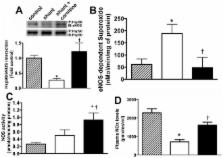
          In our model of congenital heart disease (CHD) with increased pulmonary blood flow (Shunt), we have recently shown a disruption in carnitine homeostasis, associated with mitochondrial dysfunction and decreased eNOS/Hsp90 interactions that contribute to eNOS uncoupling, increased superoxide levels, and decreased bioavailable NO. Thus, we undertook this study to test the hypothesis that L-carnitine therapy would maintain mitochondrial function, and NO signaling.

          Methods

          Thirteen fetal lambs underwent in utero placement of an aortopulmonary graft. Immediately following delivery, lambs received daily treatment with oral L-carnitine or its vehicle.

          Results

          L-carnitine-treated lambs had decreased levels of acyl carnitine, and a reduced acyl carnitine: free carnitine ratio compared to vehicle treated Shunt lambs. These changes correlated with increased carnitine acetyl transferase (CrAT) protein and enzyme activity and decreased levels of nitrated CrAT. The lactate: pyruvate ratio was also decreased in L-carnitine-treated lambs. Hsp70 protein levels were significantly decreased and this correlated with increases in eNOS/Hsp90 interactions, NOS activity, NOx levels, and a significant decrease in eNOS-derived superoxide. Further, acetylcholine significantly decreased left pulmonary vascular resistance (PVR) only in L-carnitine-treated lambs.

          Conclusion

          L-carnitine therapy may improve the endothelial dysfunction noted in children with CHD, and has important clinical implications that warrant further investigation.

          Related collections

          Most cited references42

          • Record: found
          • Abstract: found
          • Article: not found

          Mitochondrial reactive oxygen species-mediated signaling in endothelial cells.

          Once thought of as toxic by-products of cellular metabolism, reactive oxygen species (ROS) have been implicated in a large variety of cell-signaling processes. Several enzymatic systems contribute to ROS production in vascular endothelial cells, including NA(D)PH oxidase, xanthine oxidase, uncoupled endothelial nitric oxide synthase, and the mitochondrial electron transport chain. The respiratory chain is the major source of ROS in most mammalian cells, but the role of mitochondria-derived ROS in vascular cell signaling has received little attention. A new paradigm has evolved in recent years postulating that, in addition to producing ATP, mitochondria also play a key role in cell signaling and regulate a variety of cellular functions. This review focuses on the emerging role of mitochondrial ROS as signaling molecules in vascular endothelial cells. Specifically, we discuss some recent findings that indicate that mitochondrial ROS regulate vascular endothelial function, focusing on major sites of ROS production in endothelial mitochondria, factors modulating mitochondrial ROS production, the physiological and clinical implications of endothelial mitochondrial ROS, and methodological considerations in the study of mitochondrial contribution to vascular ROS generation.
            Bookmark
            • Record: found
            • Abstract: found
            • Article: not found

            Mitochondrial dysfunction, free radical generation and cellular stress response in neurodegenerative disorders.

            Protein conformational diseases, such as Alzheimer's, Parkinson's and Huntington's, affect a large portion of aging population. The pathogenic dysfunctional aggregation of proteins in non-native conformations is associated with metabolic derangements and excessive production of reactive oxygen species. Reduction of cellular expression and activity of antioxidant proteins result in increased oxidative stress. Free-radicals derived from mitochondrial dysfunction and from the cyclooxygenase enzyme activity play a role in oxidative damage of brain. Cyclooxygenase also mediates in neuro-inflammation by the production of pro-inflammatory prostaglandins which contribute to brain injury. The pathogenic role of cyclooxygenase has been demonstrated in Alzheimer and Parkinson diseases. The brain responses to detect and control diverse forms of stress are accomplished by a complex network of "longevity assurance processes" integrated to the expression of genes termed vitagenes. Heat shock proteins are a highly conserved system responsible for the preservation and repair of correct protein conformation. Heme oxygenase-1, a inducible and redox-regulated enzyme, is currently considered as having an important role in cellular antioxidant defense. A neuroprotective effect, due to its heme degrading activity, and tissue-specific pro-oxidant effects, due to its products CO and free iron, are under debate. There is a current interest in dietary compounds that can inhibit, retard or reverse the multi-stage pathophysiology of Alzheimer disease, with a chronic inflammatory response, brain injury and beta-amyloid associated pathology. Curcumin and ferulic acid, two powerful antioxidants, the first from the curry spice turmeric and the second a major constituent of fruit and vegetables, have emerged as strong inducers of the heat shock response. Food supplementation with curcumin and ferulic acid is considered a nutritional approach to reduce oxidative damage and amyloid pathology in Alzheimer disease.
              Bookmark
              • Record: found
              • Abstract: found
              • Article: not found

              Pharmacokinetics of L-carnitine.

              L-Carnitine is a naturally occurring compound that facilitates the transport of fatty acids into mitochondria for beta-oxidation. Exogenous L-carnitine is used clinically for the treatment of carnitine deficiency disorders and a range of other conditions. In humans, the endogenous carnitine pool, which comprises free L-carnitine and a range of short-, medium- and long-chain esters, is maintained by absorption of L-carnitine from dietary sources, biosynthesis within the body and extensive renal tubular reabsorption from glomerular filtrate. In addition, carrier-mediated transport ensures high tissue-to-plasma concentration ratios in tissues that depend critically on fatty acid oxidation. The absorption of L-carnitine after oral administration occurs partly via carrier-mediated transport and partly by passive diffusion. After oral doses of 1-6g, the absolute bioavailability is 5-18%. In contrast, the bioavailability of dietary L-carnitine may be as high as 75%. Therefore, pharmacological or supplemental doses of L-carnitine are absorbed less efficiently than the relatively smaller amounts present within a normal diet.L-Carnitine and its short-chain esters do not bind to plasma proteins and, although blood cells contain L-carnitine, the rate of distribution between erythrocytes and plasma is extremely slow in whole blood. After intravenous administration, the initial distribution volume of L-carnitine is typically about 0.2-0.3 L/kg, which corresponds to extracellular fluid volume. There are at least three distinct pharmacokinetic compartments for L-carnitine, with the slowest equilibrating pool comprising skeletal and cardiac muscle.L-Carnitine is eliminated from the body mainly via urinary excretion. Under baseline conditions, the renal clearance of L-carnitine (1-3 mL/min) is substantially less than glomerular filtration rate (GFR), indicating extensive (98-99%) tubular reabsorption. The threshold concentration for tubular reabsorption (above which the fractional reabsorption begins to decline) is about 40-60 micromol/L, which is similar to the endogenous plasma L-carnitine level. Therefore, the renal clearance of L-carnitine increases after exogenous administration, approaching GFR after high intravenous doses. Patients with primary carnitine deficiency display alterations in the renal handling of L-carnitine and/or the transport of the compound into muscle tissue. Similarly, many forms of secondary carnitine deficiency, including some drug-induced disorders, arise from impaired renal tubular reabsorption. Patients with end-stage renal disease undergoing dialysis can develop a secondary carnitine deficiency due to the unrestricted loss of L-carnitine through the dialyser, and L-carnitine has been used for treatment of some patients during long-term haemodialysis. Recent studies have started to shed light on the pharmacokinetics of L-carnitine when used in haemodialysis patients.
                Bookmark

                Author and article information

                Journal
                0100714
                6400
                Pediatr Res
                Pediatr. Res.
                Pediatric research
                0031-3998
                1530-0447
                1 April 2013
                29 April 2013
                July 2013
                01 January 2014
                : 74
                : 1
                : 39-47
                Affiliations
                [1 ]Pulmonary Vascular Disease Program, Vascular Biology Center, Georgia Health Sciences University, Augusta GA 30912
                [2 ]Department of Pediatrics, University of California, San Francisco CA
                [3 ]Department of Pediatrics, University Autonomous Barcelona, Spain
                [4 ]Cardiovascular Research Institute, University of California, San Francisco CA
                [5 ]Department of Cardiothoracic Surgery, University of California, Davis CA
                [6 ]Department of Pediatric Cardiology and Congenital Heart Disease, Deutsches Herzzentrum München, Klinik an der Technischen Universität München, Lazarettstrasse 36, 80636 Munich, Germany
                Author notes
                Please address correspondence and proofs to: Stephen Black, Vascular Biology Center, 1459 Laney Walker Blvd, CB3210B, Georgia Health Sciences University Augusta, GA 30912
                [*]

                These authors contributed equally

                Article
                NIHMS447492
                10.1038/pr.2013.71
                3709010
                23628882
                de58cfed-4a64-450b-8753-de69295a15e4

                Users may view, print, copy, download and text and data- mine the content in such documents, for the purposes of academic research, subject always to the full Conditions of use: http://www.nature.com/authors/editorial_policies/license.html#terms

                History
                Funding
                Funded by: National Heart, Lung, and Blood Institute : NHLBI
                Award ID: K08 HL086513 || HL
                Categories
                Article

                Pediatrics
                Pediatrics

                Comments

                Comment on this article