7
views
0
recommends
+1 Recommend
0 collections
    0
    shares
      • Record: found
      • Abstract: found
      • Article: found
      Is Open Access

      Ependymal Cilia: Physiology and Role in Hydrocephalus

      review-article

      Read this article at

      Bookmark
          There is no author summary for this article yet. Authors can add summaries to their articles on ScienceOpen to make them more accessible to a non-specialist audience.

          Abstract

          Cerebrospinal fluid (CSF), a colorless liquid that generally circulates from the lateral ventricles to the third and fourth ventricles, provides essential nutrients for brain homeostasis and growth factors during development. As evidenced by an increasing corpus of research, CSF serves a range of important functions. While it is considered that decreased CSF flow is associated to the development of hydrocephalus, it has recently been postulated that motile cilia, which line the apical surfaces of ependymal cells (ECs), play a role in stimulating CSF circulation by cilia beating. Ependymal cilia protrude from ECs, and their synchronous pulsing transports CSF from the lateral ventricle to the third and fourth ventricles, and then to the subarachnoid cavity for absorption. As a result, we postulated that malfunctioning ependymal cilia could disrupt normal CSF flow, raising the risk of hydrocephalus. This review aims to demonstrate the physiological functions of ependymal cilia, as well as how cilia immobility or disorientation causes problems. We also conclude conceivable ways of treatment of hydrocephalus currently for clinical application and provide theoretical support for regimen improvements by investigating the relationship between ependymal cilia and hydrocephalus development.

          Related collections

          Most cited references98

          • Record: found
          • Abstract: found
          • Article: found
          Is Open Access

          Multiplicity of cerebrospinal fluid functions: New challenges in health and disease

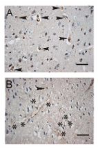
          This review integrates eight aspects of cerebrospinal fluid (CSF) circulatory dynamics: formation rate, pressure, flow, volume, turnover rate, composition, recycling and reabsorption. Novel ways to modulate CSF formation emanate from recent analyses of choroid plexus transcription factors (E2F5), ion transporters (NaHCO3 cotransport), transport enzymes (isoforms of carbonic anhydrase), aquaporin 1 regulation, and plasticity of receptors for fluid-regulating neuropeptides. A greater appreciation of CSF pressure (CSFP) is being generated by fresh insights on peptidergic regulatory servomechanisms, the role of dysfunctional ependyma and circumventricular organs in causing congenital hydrocephalus, and the clinical use of algorithms to delineate CSFP waveforms for diagnostic and prognostic utility. Increasing attention focuses on CSF flow: how it impacts cerebral metabolism and hemodynamics, neural stem cell progression in the subventricular zone, and catabolite/peptide clearance from the CNS. The pathophysiological significance of changes in CSF volume is assessed from the respective viewpoints of hemodynamics (choroid plexus blood flow and pulsatility), hydrodynamics (choroidal hypo- and hypersecretion) and neuroendocrine factors (i.e., coordinated regulation by atrial natriuretic peptide, arginine vasopressin and basic fibroblast growth factor). In aging, normal pressure hydrocephalus and Alzheimer's disease, the expanding CSF space reduces the CSF turnover rate, thus compromising the CSF sink action to clear harmful metabolites (e.g., amyloid) from the CNS. Dwindling CSF dynamics greatly harms the interstitial environment of neurons. Accordingly the altered CSF composition in neurodegenerative diseases and senescence, because of adverse effects on neural processes and cognition, needs more effective clinical management. CSF recycling between subarachnoid space, brain and ventricles promotes interstitial fluid (ISF) convection with both trophic and excretory benefits. Finally, CSF reabsorption via multiple pathways (olfactory and spinal arachnoidal bulk flow) is likely complemented by fluid clearance across capillary walls (aquaporin 4) and arachnoid villi when CSFP and fluid retention are markedly elevated. A model is presented that links CSF and ISF homeostasis to coordinated fluxes of water and solutes at both the blood-CSF and blood-brain transport interfaces. Outline 1 Overview 2 CSF formation 2.1 Transcription factors 2.2 Ion transporters 2.3 Enzymes that modulate transport 2.4 Aquaporins or water channels 2.5 Receptors for neuropeptides 3 CSF pressure 3.1 Servomechanism regulatory hypothesis 3.2 Ontogeny of CSF pressure generation 3.3 Congenital hydrocephalus and periventricular regions 3.4 Brain response to elevated CSF pressure 3.5 Advances in measuring CSF waveforms 4 CSF flow 4.1 CSF flow and brain metabolism 4.2 Flow effects on fetal germinal matrix 4.3 Decreasing CSF flow in aging CNS 4.4 Refinement of non-invasive flow measurements 5 CSF volume 5.1 Hemodynamic factors 5.2 Hydrodynamic factors 5.3 Neuroendocrine factors 6 CSF turnover rate 6.1 Adverse effect of ventriculomegaly 6.2 Attenuated CSF sink action 7 CSF composition 7.1 Kidney-like action of CP-CSF system 7.2 Altered CSF biochemistry in aging and disease 7.3 Importance of clearance transport 7.4 Therapeutic manipulation of composition 8 CSF recycling in relation to ISF dynamics 8.1 CSF exchange with brain interstitium 8.2 Components of ISF movement in brain 8.3 Compromised ISF/CSF dynamics and amyloid retention 9 CSF reabsorption 9.1 Arachnoidal outflow resistance 9.2 Arachnoid villi vs. olfactory drainage routes 9.3 Fluid reabsorption along spinal nerves 9.4 Reabsorption across capillary aquaporin channels 10 Developing translationally effective models for restoring CSF balance 11 Conclusion
            Bookmark
            • Record: found
            • Abstract: found
            • Article: not found

            New neurons follow the flow of cerebrospinal fluid in the adult brain.

            In the adult brain, neuroblasts born in the subventricular zone migrate from the walls of the lateral ventricles to the olfactory bulb. How do these cells orient over such a long distance and through complex territories? Here we show that neuroblast migration parallels cerebrospinal fluid (CSF) flow. Beating of ependymal cilia is required for normal CSF flow, concentration gradient formation of CSF guidance molecules, and directional migration of neuroblasts. Results suggest that polarized epithelial cells contribute important vectorial information for guidance of young, migrating neurons.
              Bookmark
              • Record: found
              • Abstract: found
              • Article: found

              Motile and non-motile cilia in human pathology: from function to phenotypes.

              Ciliopathies are inherited human disorders caused by both motile and non-motile cilia dysfunction that form an important and rapidly expanding disease category. Ciliopathies are complex conditions to diagnose, being multisystem disorders characterized by extensive genetic heterogeneity and clinical variability with high levels of lethality. There is marked phenotypic overlap among distinct ciliopathy syndromes that presents a major challenge for their recognition, diagnosis, and clinical management, in addition to posing an on-going task to develop the most appropriate family counselling. The impact of next-generation sequencing and high-throughput technologies in the last decade has significantly improved our understanding of the biological basis of ciliopathy disorders, enhancing our ability to determine the possible reasons for the extensive overlap in their symptoms and genetic aetiologies. Here, we review the diverse functions of cilia in human health and disease and discuss a growing shift away from the classical clinical definitions of ciliopathy syndromes to a more functional categorization. This approach arises from our improved understanding of this unique organelle, revealed through new genetic and cell biological insights into the discrete functioning of subcompartments of the cilium (basal body, transition zone, intraflagellar transport, motility). Mutations affecting these distinct ciliary protein modules can confer different genetic diseases and new clinical classifications are possible to define, according to the nature and extent of organ involvement. Copyright © 2016 Pathological Society of Great Britain and Ireland. Published by John Wiley & Sons, Ltd.
                Bookmark

                Author and article information

                Contributors
                Journal
                Front Mol Neurosci
                Front Mol Neurosci
                Front. Mol. Neurosci.
                Frontiers in Molecular Neuroscience
                Frontiers Media S.A.
                1662-5099
                12 July 2022
                2022
                : 15
                : 927479
                Affiliations
                [1] 1Department of Neurosurgery, Hunan Cancer Hospital and the Affiliated Cancer Hospital of Xiangya School of Medicine, Central South University , Changsha, China
                [2] 2Department of Neurosurgery, Xiangya Hospital, Central South University , Changsha, China
                [3] 3Diagnosis and Treatment Center for Hydrocephalus, Xiangya Hospital, Central South University , Changsha, China
                [4] 4National Clinical Research Center for Geriatric Disorders, Xiangya Hospital, Central South University , Changsha, China
                Author notes

                Edited by: Adjanie Patabendige, Edge Hill University, United Kingdom

                Reviewed by: Lance Lee, Sanford Research, United States; Ville Leinonen, Kuopio University Hospital, Finland

                *Correspondence: Gelei Xiao, xiaogelei@ 123456csu.edu.cn

                These authors have contributed equally to this work and share first authorship

                This article was submitted to Brain Disease Mechanisms, a section of the journal Frontiers in Molecular Neuroscience

                Article
                10.3389/fnmol.2022.927479
                9315228
                35903173
                e0e697db-f1b5-461d-87ac-808b0c8bad31
                Copyright © 2022 Ji, Tang, Chen, Wang, Tan, Liao, Tong and Xiao.

                This is an open-access article distributed under the terms of the Creative Commons Attribution License (CC BY). The use, distribution or reproduction in other forums is permitted, provided the original author(s) and the copyright owner(s) are credited and that the original publication in this journal is cited, in accordance with accepted academic practice. No use, distribution or reproduction is permitted which does not comply with these terms.

                History
                : 24 April 2022
                : 20 June 2022
                Page count
                Figures: 3, Tables: 1, Equations: 0, References: 98, Pages: 12, Words: 9681
                Categories
                Neuroscience
                Review

                Neurosciences
                ependymal cilia,hydrocephalus,cerebrospinal fluid,pathogenesis,treatment
                Neurosciences
                ependymal cilia, hydrocephalus, cerebrospinal fluid, pathogenesis, treatment

                Comments

                Comment on this article