- Record: found
- Abstract: found
- Article: found
mRNA Detection with Fluorescence-base Imaging Techniques for Arthritis Diagnosis
research-article
16 July 2019
Read this article at
There is no author summary for this article yet. Authors can add summaries to their articles on ScienceOpen to make them more accessible to a non-specialist audience.
Abstract
INTRODUCTION
Arthritis is a term used to indicate joint pain that are caused by inflammation of
joints or joint disease that occur commonly among older people. People of all ages,
sexes and race can and do have arthritis as well. There are different types of arthritis
with different causes and treatment methods but the most commonly known are the Rheumatoid
Arthritis (RA) and the Osteoarthritis (OA). The symptoms of arthritis are joint pain,
swelling, stiffness and decreased range of motion which could develop over time or
appear suddenly. These symptoms may or may not exacerbate over time. As the disease
get more severe, it could prevent patients from doing daily activities such as walking
up the stairs and experience chronic pain [1].
There are multiple approaches for diagnosing arthritis other than performing physical
examinations by the doctor or rheumatologists. Conventional radiography (CR) is one
of the many imaging methods for diagnosis. CR is cheap compared to the alternative,
magnetic resonance imaging (MRI) and faster than ultrasound. However, CR can only
be used towards the end of the disease process, where the bones have eroded and joint
spaces narrowing [1] and it does not show soft tissue sufficiently [2]. Both ultrasound
and MRI are not favorable due to the time it takes for diagnosis with ultrasound and
the high cost in exchange for high sensitivity of MRI. Positron emission tomography
(PET) has made its way into diagnosing the disease for its high sensitivity detection
and potential for whole-body evaluation [3]. However, due to the short half-life of
some isotopes used for this technique, patients will mostly be subjected to radiation
exposure which may cause more health problems in the future. Optical imaging (OI)
have been around in the microscopic world is now making its way to the macroscopic
setting where this technique offers high sensitivity for detection of inflammation
[4,5], fast and inexpensive and most importantly do not expose patients to ionizing
radiation [3]. OI is perfect for detecting and diagnosing rheumatoid arthritis that
mainly affects small joints of hands and feet. Many medical equipment, such as the
X-ray radiography, computed tomography (X-RAY CT), radionuclide imaging using single
photons (Single Photon Emission Computer Tomography SPECT) and positrons (Positron
Emission Tomography PET), magnetic resonance imaging (MRI), ultrasonography (US) and
optical imaging require ‘contrast agents’ for the extraction of information of the
human bodies [6,7]. Real-time PCR, fluorescence in situ hybridization (FISH) analysis
have been widely used at the cellular level to detect expression levels and cell distribution
of mRNA [8].
Examples of contrast agents are the super paramagnetic or paramagnetic metals, such
as Gadolinium Contrast Medium, used in MRI that mainly observed the whole-body scan.
To get a more target specific imaging, molecular imaging such as PET and optical imaging
like fluorescence microscopy are used for their sensitivity and specificity for target
detection. However, the disadvantages uses of PET are that they have poor spatial
and temporal resolution. They also require radioactive compounds that have intrinsically
limited half-life which limit their repeated use due to safety regulation [9,10].
In this review, we will focus on detecting mRNA at real-time with fluorescence microscopy
and the diagnosis of arthritis with fluorescence-based imaging techniques.
FLUORESCENCE-BASED IMAGING TECHNIQUES
In the past few centuries, there was a surge in the use of fluorescent dye in various
areas. Special dyes are used for printing valuable documents such as money, and certificates.
Some dyes are used to aid in leak detections of oils and fluids in vehicles. Because
of its distinct ability, the dyes are also used to assist in exploration of water
underground. Using similar concept in large scale, researchers started to apply this
approach in in vivo medical imaging. Despite countless of organic dyes in the market
for research, there are only two fluorophores that’s approved by the US Food and Drug
Administration (FDA) for medical use, which are the fluorophores are indocyanine green
(ICG) and fluorescein [11–13]. ICG is used as an ophthalmologic agent and as a hepatic
functional agent while fluorescein is used primarily in ophthalmology. These two agents,
with no toxicity reported, are primarily used to obtained retinal angiograms that
require high dosage of the agents. Even though fluorophores dyes in optical imaging
have made great strides in the imaging field, an ideal labeling reagent would be the
ones that remain non-fluorescent until bound to its target.
Fluorescence microscopy, providing real-time visualization in the surgical field,
has similar sensitivity to radionuclide imaging, providing high resolution, high contrast,
high specificity and quantitative of the sample. The downside of optical imaging is
that uncertainty emerges as the scans get into deep tissue due to unpredictable light
scattering and absorption [14,15]. Hence, the best place to use optical imaging is
on the superficial tissue surfaces such as the breasts or the lymph nodes [16–20].
However, with the continued growth in the medical imaging techniques, dyes used for
contrast images have improved significantly.
To obtain a successful optical molecular probe for medical imaging, the right wavelength
must be employed to excite the dye to obtain a unique emission wavelength. However,
if the excitation wavelength is near the ultraviolet region, the tissue will be damaged,
while excitation on at the infrared region will cause tissue heating. Absorbance and
auto fluorescence could be observed in any case when there is asymmetrical stokes
shift [12]. To prevent the auto fluorescence by the tissue under low interference,
many researchers and scientists have started applying light in the near-infra red
(NIR) wavelength (650–900 nm) as the excitation energy. These NIR wavelengths are
invisible to human eyes [21]. The incorporation of NIR fluorophores with current optical
imaging techniques provides an intraoperative image-guided surgery that allows for
target-specific imaging due to ultralow background auto fluorescence [15].
Previous study of OI have shown promising results after injection of fluorescent dyes
for detection of arthritis [4,5,22,23]. Another study has successfully shown that
in vivo leukocyte cell can be tracked by using optical imaging techniques. These in
vivo leukocyte cells are labeled with fluorescent dyes, which are suitable for the
detection of synovial inflammation in an antigen-induced arthritis model [3].
There are few available fluorescence-based imaging techniques: fluorescence microscopy,
flow cytometry, cell sorting fluorescence correlation spectroscopy as well as particle
tracking velocimetry [7].
Visualization mRNA with different hybridization methods
FISH
The localization of mRNA in cell has been around since 1980s, but then, mRNA translation
could only be visualized via in situ hybridization (ISH) using probes with multiple
fluorophores or multiple probes with one fluorophore [24]. The groups have succeeded
in transcript detection in situ, enabling genotyping of individual transcript molecule.
The earliest single molecular RNA imaging studies was transcribed and fluorescently
labeled in vitro [25]. As seen in Figure 1A, hybridization-based methods for RNA imaging
are as shown. Fluorescence in situ Hybridization (FISH) can differentiate RNA molecules
with just a single base because it only binds to specific parts of the nucleic acid.
FISH are convenient procedures due to its highly sequence-specific when combined with
amplifications during procedures in fixed cells [26,27]. Not only FISH can detect
and localized RNA targets in cells, cancer cells and tissues, it also assists in defining
dimensions of gene expression in cells and tissues in addition to exploring cell reproduction
cycle. The fluorophores are usually attached to a strand analogous to its target and
in FISH, the progress of the probes can be observed from the very beginning to the
end, when it reaches its target, because of its fluorescence nature. It is also important
to note that the probe size must be small enough to complement the target, and that
multiple colored probes (with different wavelengths) can be used together.
Molecular beacons
Molecular beacons (MB) have been used in variety of applications involving real-time
mRNA detection in living cells [28], DNA-RNA hybridization studies and protein/DNA
interactions [29]. MB, as seen in Figure 2, is a hairpin-like oligonucleotides probe
consisted of single-stranded DNA molecule in a stem-loop conformation with a fluorophore
linked to 5’ end and a quencher at the 3’ end with a minimal distance from each other.
When the molecular beacon reaches its target, the hairpin-like structure then stretches
out, separating the fluorophore and quencher, enabling fluorescence, with about 100
times more intensely than background levels of unbound probes.
An example for the success of molecular beacon is explained in a paper published in
2011 by Bratu et al. [29]. Molecular beacons in the paper are synthesized from modified
nucleic acids (2’-O-methyl RNA and DNA) to increase target specificity and sensitivity
and labeled with various fluorophores and specific quenchers respectively as seen
in Tables 1 and 2. They concluded that they were able to synthesize small MB from
2’-O-methyl RNA/LNA chimeric nucleic acids and these hairpins are observed to be stable
in cellular environment and high affinity for binding to target RNAs. They also demonstrated
that different fluorophores can be used to make the tiny MB which aided in detecting
highly structured RNAs, small RNAs and microRNAs which can be seen in Tables 1 and
2.
Upon the success of molecular beacons for fluorescence optical imaging, another research
group have developed ratiometric bimolecular beacons (RBMBs), to scan the directed
transport of single engineered RNA transcripts in living cells in real-time in 2013.
These RBMBs are designed to overcome the hurdles posed by conventional MBs and to
improve signal-to-background ratio [30]. When unbound to the target, RBMBs are in
a resting state, which is the hairpin structure like MBs. However, RBMBs do not have
fluorophore in the 5’ end and quencher in the 3’ end. As seen in Figure 3, an extra
unquenched reference dye was introduced in the 5’ end of the 18-base pair double-stranded
domain beacon leaving the 3’UU end unhinges. The fluorophore and quencher are right
next to each other, the uncertainty of the distances between fluorophores and quenchers
can be eliminated in this design.
This design was hypothesized to reduce level of false-positive detected for 24 h compared
to the conventional MBs. The unquenched reference dye is acting like a control to
allow measurements of the reporter to be adjusted for differences in RBMBs delivery
leading to more precise measurement of RNA hybridization. Their conclusion in this
study that RBMBs may be the tools to analyze single engineered RNA transcripts in
living cells.
FIT
A different approach to increase fluorescent signal upon bonding is to use forced
intercalation (FIT) probes as seen in Figure 1C. Like FISH, FIT probes are made of
peptide nucleic acid (PNA) or DNA single strands that only fluoresce as soon as it
hits the target DNA [31,32]. This is not covered in the review.
QUANTUM DOTS
Quantum dots (QD) are a new class of fluorescent probes starting to emerge in the
past few decades. QDs also have long lifetime increasing the probability of adsorption
and producing broad absorption spectrum. Other than being non-toxic to samples, one
of the most appreciated advantages of QD is their photostability or resistance to
photobleaching, which allows images to be recorded over a longer period compared to
conventional fluorescent dyes [7]. Other than in vivo targeting and imaging, QDs are
also used in cellular targeting and imaging, as well as fixed tissue analysis, optical
encoding and quantitative determination [33].
Size of the quantum dot nanoparticles in bioimaging field generally falls within 2–10
nm in diameter. QDs have unique optical and electronic properties, with molar extinction
coefficients that are 10–50 times larger, making it much brighter than conventional
dyes. The emission wavelengths are size tunable depending on the radius of the QD.
Larger QDs were also synthesized to be used in other applications other than in vivo
imaging. However, parameters such as the distance between gold nanoparticle to optical
absorbance and scattering intensity of the inter-particle distance can be controlled,
resulting in a research group utilizing 20 nm Au nanoparticles QD to predict the localized
surface plasmon resonance (LSPR) of gold nanoparticles. They were able to synthesize
20 nm AuNP monomers with zero LSPR background as probes with only a single target
molecule required to form a dimer with significant plasmon resonance coupling effect,
increasing the sensitivity of the LSPR sensor to single cell level. In their study,
they were able to present a smart single mRNA imaging approach in living cells based
on target-induced formation of nanoparticle dimers. They concluded that with proper
linker, these nanoparticles could serve as a basis to many imaging techniques for
biomolecules and mRNA in living cells [34].
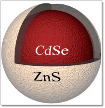
The optical properties of quantum dots originated from their elemental composition,
with a semiconductor core such as cadmium selenide (CdSe) or lead selenide (PbSe),
coated with a semiconductor shell, as seen in Figure 4. Properties of QDs have made
an impression in the optical imaging field, prompted a topic of intensive interest
in cancer biology, molecular imaging and molecular profiling [32,35–42]. Although
QDs presented the researchers numerous advantages over traditional organic dyes, these
inorganic semiconductor materials are toxic to living systems, limiting their use
in biological systems, triggering a hot topic for research in the field on synthesizing
a more biocompatible QD for the biological systems. Multiples experiments have been
conducted on modified QDs, such as extra surface coating to minimize the cytotoxicity
[32]. For example, cadmium ions present in the many QDs are shown to bind to thiol
groups on important molecules in the mitochondria causing cell death [32]. The cadmium
ions present in the cells are most likely caused by QD exposure to air and UV light,
causing the surface to oxidize in oxidative solution. The levels of cytotoxicity could
be reduced or eliminated by addition surface coatings [32].
QUANTUM DOTS and MOLECULAR BEACONS
Since quantum dots have become interesting fluorophores for biological imaging fields,
researchers have developed a new method for the fluorescence imaging of endogenous
mRNA using signal-tunable molecular beacon technique based on QDs in living cells.
As seen in Figure 5, the group targeted mRNA124a which was observed to have high expression
during neuronal development [43]. The beacons, each composed of a quencher on the
mRNA124a binding sequence and the R9 peptide, are incorporated with a quantum dots
to form R9-QD-mRNA124a beacon. In the absence of mRNA124a, R9-QD-mRNA124a formed a
partial duplex beacon that quenches the system, giving no fluorescence signal. With
mRNA124a, the mRNA124a binding sequences leave together with the quenchers, leaving
a signal of red fluorescence. They concluded that the method could provide critical
information on expression during neurogenesis and could be applied to various systems
to track cellular developments with different sized QD-based molecular system.
CONCLUSION
There are many restrictions obtaining multicolor in vivo imaging mainly due to overlapping
fluorescence emissions. Different approaches have been executed, including the recently
developed 3’untranslated region (UTR)-based reported gene imaging system to monitor
the expression of patterns of mRNA. However, with this approach, it is difficult to
differentiate if the data obtained are from the mRNA expression or from cell death
in vivo [44–47].
On the other hand, different fluorophores with discernable emissions wavelength must
be chosen for multicolor imaging. The complications for the traditional dyes such
as organic dyes and QDs are the requirement to use UV or short-wavelength radiation
for excitation of the materials and their cytotoxicity in biological environment.
Low light penetration depth is observed due to the short wavelength excitation light
or low signal-to-noise ratio due to auto-fluorescence. Although fluorescence-based
imaging techniques have improved tremendously, there are still rooms to improve the
techniques or the probes. It is important to develop more efficient bio-labels to
overcome these limitations. NIR probes have gained its momentum the past decades to
overcome the shortcomings of the traditional probes when it comes to monitoring the
sample with higher depth. Few companies have developed some NIR fluorophores with
emission >800 nm, permitting NIR probes to be used together with the original fluorescent
organic dye [48]. With the developments of new types of contrast agents together with
improvement in electronics and software in equipment, fluorescence-based imaging technique
can expand even more. In conjunction of NIR probes, quantum dots have also been an
interesting tool not only for imaging field, but for the computing as well as the
quantum dot displays for lightning up LCD displays in televisions. Many researches
could be done in terms of quantum dots in the field of nanotechnology. Future work
such as incorporating the reporter tag into endogenous gene loci using CRISPRCas0
genome editing tools would proof to be quite interesting [49].
The NIR dye, ICG, is already being used to test for liver cancer during surgery, to
identify small and unidentifiable liver cancers in real time. ICG has also been used
to identify lymph nodes in real time in a small number of breast cancer patients [50].
Even though optical imaging has yet to replace conventional imaging as a form of arthritis
tool, OI is a promising diagnostic tool for detecting early onset arthritis.
In summary, this review analysis on the visualization of mRNA with different hybridization
techniques. There were many shortcomings since the development of fluorescence-based
imaging techniques, but scientists and researchers have all overcome the obstacles
and improved both the probes and instruments for a better future. The capability of
fluorescence-based imaging techniques is open to new possibilities for better and
faster diagnostic of joint diseases in the future.
Related collections
Most cited references49
- Record: found
- Abstract: found
- Article: not found
Development and applications of CRISPR-Cas9 for genome engineering.
Patrick D Hsu, Eric S. Lander, Feng Zhang (2014)
- Record: found
- Abstract: not found
- Article: not found
New strategies for fluorescent probe design in medical diagnostic imaging.
Hisataka Kobayashi, Mikako Ogawa, Raphael Alford … (2010)
- Record: found
- Abstract: found
- Article: not found
In vivo imaging of tumors with protease-activated near-infrared fluorescent probes.
R Weissleder, C. H. Tung, U Mahmood … (1999)