21
views
0
recommends
+1 Recommend
0 collections
    0
    shares
      • Record: found
      • Abstract: found
      • Article: found
      Is Open Access

      Cherubism: A rare case report

      case-report

      Read this article at

      Bookmark
          There is no author summary for this article yet. Authors can add summaries to their articles on ScienceOpen to make them more accessible to a non-specialist audience.

          Abstract

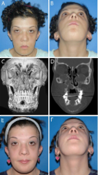
          Cherubism is a rare congenital disease resulting in malformation of the jaw. It occurs before the age of 5 years and regress spontaneously after puberty. It can result into enlargement of the jaw bone, tooth displacement, facial disfigurement and psychological trauma to patient. Hence, the understanding about the condition, its progression and management is necessary.

          Related collections

          Most cited references14

          • Record: found
          • Abstract: found
          • Article: found
          Is Open Access

          Cherubism: best clinical practice

          Cherubism is a skeletal dysplasia characterized by bilateral and symmetric fibro-osseous lesions limited to the mandible and maxilla. In most patients, cherubism is due to dominant mutations in the SH3BP2 gene on chromosome 4p16.3. Affected children appear normal at birth. Swelling of the jaws usually appears between 2 and 7 years of age, after which, lesions proliferate and increase in size until puberty. The lesions subsequently begin to regress, fill with bone and remodel until age 30, when they are frequently not detectable. Fibro-osseous lesions, including those in cherubism have been classified as quiescent, non-aggressive and aggressive on the basis of clinical behavior and radiographic findings. Quiescent cherubic lesions are usually seen in older patients and do not demonstrate progressive growth. Non-aggressive lesions are most frequently present in teenagers. Lesions in the aggressive form of cherubism occur in young children and are large, rapidly growing and may cause tooth displacement, root resorption, thinning and perforation of cortical bone. Because cherubism is usually self-limiting, operative treatment may not be necessary. Longitudinal observation and follow-up is the initial management in most cases. Surgical intervention with curettage, contouring or resection may be indicated for functional or aesthetic reasons. Surgical procedures are usually performed when the disease becomes quiescent. Aggressive lesions that cause severe functional problems such as airway obstruction justify early surgical intervention.
            Bookmark
            • Record: found
            • Abstract: found
            • Article: found
            Is Open Access

            Cherubism: Clinicoradiographic Features and Treatment

            ABSTRACT Objectives Cherubism is a congenital childhood disease of autosomal dominant inheritance. This disease is characterized by painless bilateral enlargement of the jaws, in which bone is replaced with fibrous tissue. The condition has sui generis clinical, radiographic and histological features, of which the clinician should be aware for a better differential diagnosis in the presence of a fibro-osseous lesion affecting the bones of the maxillomandibular complex. The purpose of present paper was to review the literature and to report the most important aspects of cherubism in order to facilitate the study of this disease Material and Methods Literature was reviewed about cherubism, emphasizing the relevant clinicoradiographic features and treatment. Literature was selected through a search of PubMed and Scielo electronic databases. The keywords used for search were adolescent, cherubism, cherubism/physiopathology, cherubism/treatment, cherubism/radiography. A manual search of the reference lists of the identified articles and the authors' article files and recent reviews was conducted to identify additional publications. Those studies that described new features about cherubism were included in this review. Results In total 44 literature sources were obtained and reviewed. Studies that described new features about cherubism physiopathology, diagnostics and treatment were reviewed. Conclusions Despite the exceptions, cherubism is a clinically well-characterized disease. In cases of a suspicion of cherubism, radiographic examination is essential since the clinical presentation, the location and distribution of the lesions may define the diagnosis. Histopathological examination is complementary. Nowadays, genetic tests should be used for final diagnosis of cherubism.
              Bookmark
              • Record: found
              • Abstract: found
              • Article: not found

              An extreme case of cherubism.

              We describe an 8-year-old boy who presented with severe facial swelling. This progressed rapidly and 17 months later he died of gastrointestinal and pulmonary infections. The diagnosis was initially brown tumour associated with hyperparathyroidism, but this was revised in the light of laboratory investigations that were within the reference ranges, and normal appearance of the parathyroids on exploration to that of an extreme case of cherubism that behaved in a locally aggressive manner. Copyright 2002 The British Association of Oral and Maxillofacial Surgeons.
                Bookmark

                Author and article information

                Journal
                J Nat Sci Biol Med
                J Nat Sci Biol Med
                JNSBM
                Journal of Natural Science, Biology, and Medicine
                Medknow Publications & Media Pvt Ltd (India )
                0976-9668
                2229-7707
                Jul-Dec 2014
                : 5
                : 2
                : 488-491
                Affiliations
                [1 ] Department of Oral Pathology, Raj Rajeswari Dental College, Udaipur, Rajasthan, India
                [2 ] Department of Oral Pathology, Raj Rajeswari Dental College, Udaipur, Rajasthan, India
                [3 ] Department of Pedodontics, Vyas Dental College, Jodhpur, Rajasthan, India
                [4 ] Department of Orthodontics, Maharana Pratap Dental College, Kanpur, Utter Pradesh, India
                [5 ] Department of Oral Pathology, Guardian Dental College, Ambernath, Maharastra, India
                [6 ] Department of Prosthodontics, MIDSR Dental College, Latur, Maharastra, India
                Author notes
                Address for correspondence: Dr. Prashant Babaji, Department of Pedodontics, Vyas Dental College, Jodhpur - 342 001, Rajasthan, India. E-mail: babajipedo@ 123456rediffmail.com
                Article
                JNSBM-5-488
                10.4103/0976-9668.136283
                4121945
                25097445
                eb13ef8a-91da-4b8f-b413-bfbe61a46930
                Copyright: © Journal of Natural Science, Biology and Medicine

                This is an open-access article distributed under the terms of the Creative Commons Attribution-Noncommercial-Share Alike 3.0 Unported, which permits unrestricted use, distribution, and reproduction in any medium, provided the original work is properly cited.

                History
                Categories
                Case Reports

                Life sciences
                cherubism,childhood disease,giant cell lesion,jaw growth,malocclusion
                Life sciences
                cherubism, childhood disease, giant cell lesion, jaw growth, malocclusion

                Comments

                Comment on this article