38
views
0
recommends
+1 Recommend
0 collections
    0
    shares
      • Record: found
      • Abstract: found
      • Article: found
      Is Open Access

      CD45 regulates retention, motility, and numbers of hematopoietic progenitors, and affects osteoclast remodeling of metaphyseal trabecules

      research-article

      Read this article at

      Bookmark
          There is no author summary for this article yet. Authors can add summaries to their articles on ScienceOpen to make them more accessible to a non-specialist audience.

          Abstract

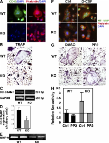
          The CD45 phosphatase is uniquely expressed by all leukocytes, but its role in regulating hematopoietic progenitors is poorly understood. We show that enhanced CD45 expression on bone marrow (BM) leukocytes correlates with increased cell motility in response to stress signals. Moreover, immature CD45 knockout (KO) cells showed defective motility, including reduced homing (both steady state and in response to stromal-derived factor 1) and reduced granulocyte colony-stimulating factor mobilization. These defects were associated with increased cell adhesion mediated by reduced matrix metalloproteinase 9 secretion and imbalanced Src kinase activity. Poor mobilization of CD45KO progenitors by the receptor activator of nuclear factor κB ligand, and impaired modulation of the endosteal components osteopontin and stem cell factor, suggested defective osteoclast function. Indeed, CD45KO osteoclasts exhibited impaired bone remodeling and abnormal morphology, which we attributed to defective cell fusion and Src function. This led to irregular distribution of metaphyseal bone trabecules, a region enriched with stem cell niches. Consequently, CD45KO mice had less primitive cells in the BM and increased numbers of these cells in the spleen, yet with reduced homing and repopulation potential. Uncoupling environmental and intrinsic defects in chimeric mice, we demonstrated that CD45 regulates progenitor movement and retention by influencing both the hematopoietic and nonhematopoietic compartments.

          Related collections

          Most cited references42

          • Record: found
          • Abstract: found
          • Article: not found

          Recruitment of stem and progenitor cells from the bone marrow niche requires MMP-9 mediated release of kit-ligand.

          Stem cells within the bone marrow (BM) exist in a quiescent state or are instructed to differentiate and mobilize to circulation following specific signals. Matrix metalloproteinase-9 (MMP-9), induced in BM cells, releases soluble Kit-ligand (sKitL), permitting the transfer of endothelial and hematopoietic stem cells (HSCs) from the quiescent to proliferative niche. BM ablation induces SDF-1, which upregulates MMP-9 expression, and causes shedding of sKitL and recruitment of c-Kit+ stem/progenitors. In MMP-9-/- mice, release of sKitL and HSC motility are impaired, resulting in failure of hematopoietic recovery and increased mortality, while exogenous sKitL restores hematopoiesis and survival after BM ablation. Release of sKitL by MMP-9 enables BM repopulating cells to translocate to a permissive vascular niche favoring differentiation and reconstitution of the stem/progenitor cell pool.
            Bookmark
            • Record: found
            • Abstract: found
            • Article: not found

            G-CSF induces stem cell mobilization by decreasing bone marrow SDF-1 and up-regulating CXCR4.

            Granulocyte colony-stimulating factor (G-CSF) induced hematopoietic stem cell mobilization is widely used for clinical transplantation; however, the mechanism is poorly understood. We report here that G-CSF induced a reduction of the chemokine stromal cell derived factor 1 (SDF-1) and an increase in its receptor CXCR4 in the bone marrow (BM), whereas their protein expression in the blood was less affected. The gradual decrease of BM SDF-1, due mostly to its degradation by neutrophil elastase, correlated with stem cell mobilization. Elastase inhibition reduced both activities. Human and murine stem cell mobilization was inhibited by neutralizing CXCR4 or SDF-1 antibodies, demonstrating SDF-1 CXCR4 signaling in cell egress. We suggest that manipulation of SDF-1 CXCR4 interactions may be a means with which to control the navigation of progenitors between the BM and blood to improve the outcome of clinical stem cell transplantation.
              Bookmark
              • Record: found
              • Abstract: found
              • Article: not found

              Direct three-dimensional morphometric analysis of human cancellous bone: microstructural data from spine, femur, iliac crest, and calcaneus.

              The appearance of cancellous bone architecture is different for various skeletal sites and various disease states. During aging and disease, plates are perforated and connecting rods are dissolved. There is a continuous shift from one structural type to the other. So traditional histomorphometric procedures, which are based on a fixed model type, will lead to questionable results. The introduction of three-dimensional (3D) measuring techniques in bone research makes it possible to capture the actual architecture of cancellous bone without assumptions of the structure type. This requires, however, new methods that make direct use of the 3D information. Within the framework of a BIOMED I project of the European Union, we analyzed a total of 260 human bone biopsies taken from five different skeletal sites (femoral head, vertebral bodies L2 and L4, iliac crest, and calcaneus) from 52 donors. The samples were measured three-dimensionally with a microcomputed tomography scanner and subsequently evaluated with both traditional indirect histomorphometric methods and newly developed direct ones. The results show significant differences between the methods and in their relation to the bone volume fraction. Based on the direct 3D analysis of human bone biopsies, it appears that samples with a lower bone mass are primarily characterized by a smaller plate-to-rod ratio, and to a lesser extent by thinner trabecular elements.
                Bookmark

                Author and article information

                Journal
                J Exp Med
                jem
                The Journal of Experimental Medicine
                The Rockefeller University Press
                0022-1007
                1540-9538
                29 September 2008
                : 205
                : 10
                : 2381-2395
                Affiliations
                [1 ]Department of Immunology and [2 ]Molecular Genetics, Weizmann Institute of Science, Rehovot 76100, Israel
                [3 ]The Laboratory of Musculoskeletal Biomechanics and Applied Anatomy, Koret School of Veterinary Medicine, Hebrew University of Jerusalem, Rehovot 76100, Israel
                Author notes

                CORRESPONDENCE Tsvee Lapidot: Tsvee.Lapidot@ 123456weizmann.ac.il

                Article
                20080072
                10.1084/jem.20080072
                2556782
                18779349
                ed69ccc2-8b19-42d3-9f72-bb5168dca5d0
                © 2008 Shivtiel et al.

                This article is distributed under the terms of an Attribution–Noncommercial–Share Alike–No Mirror Sites license for the first six months after the publication date (see http://www.jem.org/misc/terms.shtml). After six months it is available under a Creative Commons License (Attribution–Noncommercial–Share Alike 3.0 Unported license, as described at http://creativecommons.org/licenses/by-nc-sa/3.0/).

                History
                : 10 January 2008
                : 14 August 2008
                Categories
                Articles
                Article

                Medicine
                Medicine

                Comments

                Comment on this article