60
views
0
recommends
+1 Recommend
0 collections
    0
    shares
      • Record: found
      • Abstract: found
      • Article: found
      Is Open Access

      Warburg effect in chemosensitivity: Targeting lactate dehydrogenase-A re-sensitizes Taxol-resistant cancer cells to Taxol

      research-article

      Read this article at

      Bookmark
          There is no author summary for this article yet. Authors can add summaries to their articles on ScienceOpen to make them more accessible to a non-specialist audience.

          Abstract

          Background

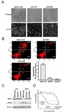
          Taxol is one of the most effective chemotherapeutic agents for the treatment of patients with breast cancer. Despite impressive clinical responses initially, the majority of patients eventually develop resistance to Taxol. Lactate dehydrogenase-A (LDH-A) is one of the predominant isoforms of LDH expressed in breast tissue, which controls the conversion of pyruvate to lactate and plays an important role in glucose metabolism. In this study we investigated the role of LDH-A in mediating Taxol resistance in human breast cancer cells.

          Results

          Taxol-resistant subclones, derived from the cancer cell line MDA-MB-435, sustained continuous growth in high concentrations of Taxol while the Taxol-sensitive cells could not. The increased expression and activity of LDH-A were detected in Taxol-resistant cells when compared with their parental cells. The downregulation of LDH-A by siRNA significantly increased the sensitivity of Taxol-resistant cells to Taxol. A higher sensitivity to the specific LDH inhibitor, oxamate, was found in the Taxol-resistant cells. Furthermore, treating cells with the combination of Taxol and oxamate showed a synergistical inhibitory effect on Taxol-resistant breast cancer cells by promoting apoptosis in these cells.

          Conclusion

          LDH-A plays an important role in Taxol resistance and inhibition of LDH-A re-sensitizes Taxol-resistant cells to Taxol. This supports that Warburg effect is a property of Taxol resistant cancer cells and may play an important role in the development of Taxol resistance. To our knowledge, this is the first report showing that the increased expression of LDH-A plays an important role in Taxol resistance of human breast cancer cells. This study provides valuable information for the future development and use of targeted therapies, such as oxamate, for the treatment of patients with Taxol-resistant breast cancer.

          Related collections

          Most cited references22

          • Record: found
          • Abstract: found
          • Article: not found

          Cancer's molecular sweet tooth and the Warburg effect.

          More than 80 years ago, the renowned biochemist Otto Warburg described how cancer cells avidly consume glucose and produce lactic acid under aerobic conditions. Recent studies arguing that cancer cells benefit from this phenomenon, termed the Warburg effect, have renewed discussions about its exact role as cause, correlate, or facilitator of cancer. Molecular advances in this area may reveal tactics to exploit the cancer cell's "sweet tooth" for cancer therapy.
            Bookmark
            • Record: found
            • Abstract: found
            • Article: not found

            Specific proteolytic cleavage of poly(ADP-ribose) polymerase: an early marker of chemotherapy-induced apoptosis.

            Apoptosis is a morphologically and biochemically distinct form of cell death that occurs under a variety of physiological and pathological conditions. In the present study, the proteolytic cleavage of poly(ADP-ribose) polymerase (pADPRp) during the course of chemotherapy-induced apoptosis was examined. Treatment of HL-60 human leukemia cells with the topoisomerase II-directed anticancer agent etoposide resulted in morphological changes characteristic of apoptosis. Endonucleolytic degradation of DNA to generate nucleosomal fragments occurred simultaneously. Western blotting with epitope-specific monoclonal and polyclonal antibodies revealed that these characteristic apoptotic changes were accompanied by early, quantitative cleavage of the M(r) 116,000 pADPRp polypeptide to an M(r) approximately 25,000 fragment containing the amino-terminal DNA-binding domain of pADPRp and an M(r) approximately 85,000 fragment containing the automodification and catalytic domains. Activity blotting revealed that the M(r) approximately 85,000 fragment retained basal pADPRp activity but was not activated by exogenous nicked DNA. Similar cleavage of pADPRp was observed after exposure of HL-60 cells to a variety of chemotherapeutic agents including cis-diaminedichloroplatinum(II), colcemid, 1-beta-D-arabinofuranosylcytosine, and methotrexate; to gamma-irradiation; or to the protein synthesis inhibitors puromycin or cycloheximide. Similar changes were observed in MDA-MB-468 human breast cancer cells treated with trifluorothymidine or 5-fluoro-2'-deoxyuridine and in gamma-irradiated or glucocorticoid-treated rat thymocytes undergoing apoptosis. Treatment with several compounds (tosyl-L-lysine chloromethyl ketone, tosyl-L-phenylalanine chloromethyl ketone, N-ethylmaleimide, iodoacetamide) prevented both the proteolytic cleavage of pADPRp and the internucleosomal fragmentation of DNA. The results suggest that proteolytic cleavage of pADPRp, in addition to being an early marker of chemotherapy-induced apoptosis, might reflect more widespread proteolysis that is a critical biochemical event early during the process of physiological cell death.
              Bookmark
              • Record: found
              • Abstract: found
              • Article: not found

              Role of poly(ADP-ribose) polymerase (PARP) cleavage in apoptosis. Caspase 3-resistant PARP mutant increases rates of apoptosis in transfected cells.

              An early transient burst of poly(ADP-ribosyl)ation of nuclear proteins was recently shown to be required for apoptosis to proceed in various cell lines (Simbulan-Rosenthal, C., Rosenthal, D., Iyer, S., Boulares, H., and Smulson, M. (1998) J. Biol. Chem. 273, 13703-13712) followed by cleavage of poly(ADP-ribose) polymerase (PARP), catalyzed by caspase-3. This inactivation of PARP has been proposed to prevent depletion of NAD (a PARP substrate) and ATP, which are thought to be required for later events in apoptosis. The role of PARP cleavage in apoptosis has now been investigated in human osteosarcoma cells and PARP -/- fibroblasts stably transfected with a vector encoding a caspase-3-resistant PARP mutant. Expression of this mutant PARP increased the rate of staurosporine and tumor necrosis factor-alpha-induced apoptosis, at least in part by reducing the time interval required for the onset of caspase-3 activation and internucleosomal DNA fragmentation, as well as the generation of 50-kilobase pair DNA breaks, thought to be associated with early chromatin unfolding. Overexpression of wild-type PARP in osteosarcoma cells also accelerated the apoptotic process, although not to the same extent as that apparent in cells expressing the mutant PARP. These effects of the mutant and wild-type enzymes might be due to the early and transient poly(ADP-ribose) synthesis in response to DNA breaks, and the accompanying depletion of NAD apparent in the transfected cells. The accelerated NAD depletion did not seem to interfere with the later stages of apoptosis. These results indicate that PARP activation and subsequent cleavage have active and complex roles in apoptosis.
                Bookmark

                Author and article information

                Journal
                Mol Cancer
                Molecular Cancer
                BioMed Central
                1476-4598
                2010
                9 February 2010
                : 9
                : 33
                Affiliations
                [1 ]Mitchell Cancer Institute, University of South Alabama, Mobile, Alabama, USA
                [2 ]Department of Cell Biology and Neuroscience, University of South Alabama, Mobile, Alabama, USA
                [3 ]Cancer Research Institute, Central South University, Changsha, China
                [4 ]Institute for Cancer Research, The Norwegian Radium Hospital, University of Oslo, Norway
                [5 ]Department of Biochemistry and Molecular Biology, University of Florida, Gainesville, Florida, USA
                [6 ]Ochsner Cancer Institute, Ochsner Health System, New Orleans, Louisiana, USA
                Article
                1476-4598-9-33
                10.1186/1476-4598-9-33
                2829492
                20144215
                edcb4c48-227a-4d65-80bb-297c0f01b65a
                Copyright ©2010 Zhou et al; licensee BioMed Central Ltd.

                This is an Open Access article distributed under the terms of the Creative Commons Attribution License ( http://creativecommons.org/licenses/by/2.0), which permits unrestricted use, distribution, and reproduction in any medium, provided the original work is properly cited.

                History
                : 15 August 2009
                : 9 February 2010
                Categories
                Research

                Oncology & Radiotherapy
                Oncology & Radiotherapy

                Comments

                Comment on this article