Inviting an author to review:
Find an author and click ‘Invite to review selected article’ near their name.
Search for authorsSearch for similar articles
64
views
0
recommends
+1 Recommend
0 collections
    0
    shares
      • Record: found
      • Abstract: found
      • Article: found
      Is Open Access

      Postextraction Alveolar Ridge Preservation: Biological Basis and Treatments

      review-article

      Read this article at

      Bookmark
          There is no author summary for this article yet. Authors can add summaries to their articles on ScienceOpen to make them more accessible to a non-specialist audience.

          Abstract

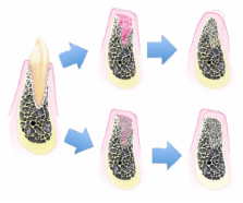
          Following tooth extraction, the alveolar ridge undergoes an inevitable remodeling process that influences implant therapy of the edentulous area. Socket grafting is a commonly adopted therapy for the preservation of alveolar bone structures in combination or not with immediate implant placement although the biological bases lying behind this treatment modality are not fully understood and often misinterpreted. This review is intended to clarify the literature support to socket grafting in order to provide practitioners with valid tools to make a conscious decision of when and why to recommend this therapy.

          Related collections

          Most cited references139

          • Record: found
          • Abstract: found
          • Article: not found

          Dimensional ridge alterations following tooth extraction. An experimental study in the dog.

          To study dimensional alterations of the alveolar ridge that occurred following tooth extraction as well as processes of bone modelling and remodelling associated with such change. Twelve mongrel dogs were included in the study. In both quadrants of the mandible incisions were made in the crevice region of the 3rd and 4th premolars. Minute buccal and lingual full thickness flaps were elevated. The four premolars were hemi-sected. The distal roots were removed. The extraction sites were covered with the mobilized gingival tissue. The extractions of the roots and the sacrifice of the dogs were staggered in such a manner that all dogs contributed with sockets representing 1, 2, 4 and 8 weeks of healing. The animals were sacrificed and tissue blocks containing the extraction socket were dissected, decalcified in EDTA, embedded in paraffin and cut in the buccal-lingual plane. The sections were stained in haematoxyline-eosine and examined in the microscope. It was demonstrated that marked dimensional alterations occurred during the first 8 weeks following the extraction of mandibular premolars. Thus, in this interval there was a marked osteoclastic activity resulting in resorption of the crestal region of both the buccal and the lingual bone wall. The reduction of the height of the walls was more pronounced at the buccal than at the lingual aspect of the extraction socket. The height reduction was accompanied by a "horizontal" bone loss that was caused by osteoclasts present in lacunae on the surface of both the buccal and the lingual bone wall. The resorption of the buccal/lingual walls of the extraction site occurred in two overlapping phases. During phase 1, the bundle bone was resorbed and replaced with woven bone. Since the crest of the buccal bone wall was comprised solely of bundle this modelling resulted in substantial vertical reduction of the buccal crest. Phase 2 included resorption that occurred from the outer surfaces of both bone walls. The reason for this additional bone loss is presently not understood. (c) Blackwell Munksgaard, 2005.
            Bookmark
            • Record: found
            • Abstract: found
            • Article: not found

            A systematic review of post-extractional alveolar hard and soft tissue dimensional changes in humans.

            Removal of teeth results in both horizontal and vertical changes of hard and soft tissue dimensions. The magnitude of these changes is important for decision-making and comprehensive treatment planning, with provisions for possible solutions to expected complications during prosthetic rehabilitation. To review all English dental literature to assess the magnitude of dimensional changes of both the hard and soft tissues of the alveolar ridge up to 12 months following tooth extraction in humans. An electronic MEDLINE and CENTRAL search complemented by manual searching was conducted to identify randomized controlled clinical trials and prospective cohort studies on hard and soft tissue dimensional changes after tooth extraction. Only studies reporting on undisturbed post-extraction dimensional changes relative to a fixed reference point over a clearly stated time period were included. Assessment of the identified studies and data extraction was performed independently by two reviewers. Data collected were reported by descriptive methods. Weighted means and percentages of the dimensional changes over time were calculated where appropriate. The search provided 3954 titles and 238 abstracts. Full text analysis was performed for 104 articles resulting in 20 studies that met the inclusion criteria. In human hard tissue, horizontal dimensional reduction (3.79 ± 0.23 mm) was more than vertical reduction (1.24 ± 0.11 mm on buccal, 0.84 ± 0.62 mm on mesial and 0.80 ± 0.71 mm on distal sites) at 6 months. Percentage vertical dimensional change was 11-22% at 6 months. Percentage horizontal dimensional change was 32% at 3 months, and 29-63% at 6-7 months. Soft tissue changes demonstrated 0.4-0.5 mm gain of thickness at 6 months on the buccal and lingual aspects. Horizontal dimensional changes of hard and soft tissue (loss of 0.1-6.1 mm) was more substantial than vertical change (loss 0.9 mm to gain 0.4 mm) during observation periods of up to 12 months, when study casts were utilized as a means of documenting the changes. Human re-entry studies showed horizontal bone loss of 29-63% and vertical bone loss of 11-22% after 6 months following tooth extraction. These studies demonstrated rapid reductions in the first 3-6 months that was followed by gradual reductions in dimensions thereafter. © 2011 John Wiley & Sons A/S.
              Bookmark
              • Record: found
              • Abstract: not found
              • Article: not found

              On the repair potential of periodontal tissues.

                Bookmark

                Author and article information

                Journal
                Int J Dent
                Int J Dent
                IJD
                International Journal of Dentistry
                Hindawi Publishing Corporation
                1687-8728
                1687-8736
                2012
                12 June 2012
                : 2012
                : 151030
                Affiliations
                1Unit of Periodontology, Department of Biomedical, Surgical and Dental Sciences, University of Milan, Foundation IRCCS Cà Granda, 20142 Milan, Italy
                2Department of Periodontics and Oral Medicine and Michigan Center for Oral Health Research, Ann Arbor, MI, USA
                3Department of Biomedical Engineering, College of Engineering, University of Michigan, Ann Arbor, MI, USA
                Author notes

                Academic Editor: Figen Cizmeci Senel

                Article
                10.1155/2012/151030
                3378971
                22737169
                f0bf8951-ff67-47e0-a017-d57e591ee3bc
                Copyright © 2012 Giorgio Pagni et al.

                This is an open access article distributed under the Creative Commons Attribution License, which permits unrestricted use, distribution, and reproduction in any medium, provided the original work is properly cited.

                History
                : 15 February 2012
                : 2 April 2012
                Categories
                Review Article

                Dentistry
                Dentistry

                Comments

                Comment on this article