24
views
0
recommends
+1 Recommend
0 collections
    0
    shares
      • Record: found
      • Abstract: found
      • Article: found
      Is Open Access

      Establishment of CMab-43, a Sensitive and Specific Anti-CD133 Monoclonal Antibody, for Immunohistochemistry

      research-article

      Read this article at

      Bookmark
          There is no author summary for this article yet. Authors can add summaries to their articles on ScienceOpen to make them more accessible to a non-specialist audience.

          Abstract

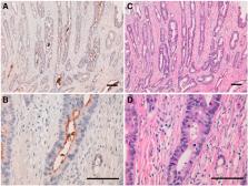
          CD133, also known as prominin-1, was first described as a cell surface marker on early progenitor and hematopoietic stem cells. It is a five-domain transmembrane protein composed of an N-terminal extracellular tail, two small cytoplasmic loops, two large extracellular loops containing seven potential glycosylation sites, and a short C-terminal intracellular tail. CD133 has been used as a marker to identify cancer stem cells derived from primary solid tumors and as a prognostic marker of gliomas. Herein, we developed novel anti-CD133 monoclonal antibodies (mAbs) and characterized their efficacy in flow cytometry, Western blot, and immunohistochemical analyses. We expressed the full length of CD133 in LN229 glioblastoma cells, immunized mice with LN229/CD133 cells, and performed the first screening using flow cytometry. After limiting dilution, we established 100 anti-CD133 mAbs, reacting with LN229/CD133 cells but not with LN229 cells. Subsequently, we performed the second and third screening with Western blot and immunohistochemical analyses, respectively. Among 100 mAbs, 11 strongly reacted with CD133 in Western blot analysis. One of 11 clones, CMab-43 (IgG 2a, kappa), showed a sensitive and specific reaction against colon cancer cells, warranting the use of CMab-43 in detecting CD133 in pathological analyses of CD133-expressing cancers.

          Related collections

          Most cited references16

          • Record: found
          • Abstract: found
          • Article: not found

          CD133(+) and CD133(-) glioblastoma-derived cancer stem cells show differential growth characteristics and molecular profiles.

          Although glioblastomas show the same histologic phenotype, biological hallmarks such as growth and differentiation properties vary considerably between individual cases. To investigate whether different subtypes of glioblastomas might originate from different cells of origin, we cultured tumor cells from 22 glioblastomas under medium conditions favoring the growth of neural and cancer stem cells (CSC). Secondary glioblastoma (n = 7)-derived cells did not show any growth in the medium used, suggesting the absence of neural stem cell-like tumor cells. In contrast, 11/15 primary glioblastomas contained a significant CD133(+) subpopulation that displayed neurosphere-like, nonadherent growth and asymmetrical cell divisions yielding cells expressing markers characteristic for all three neural lineages. Four of 15 cell lines derived from primary glioblastomas grew adherently in vitro and were driven by CD133(-) tumor cells that fulfilled stem cell criteria. Both subtypes were similarly tumorigenic in nude mice in vivo. Clinically, CD133(-) glioblastomas were characterized by a lower proliferation index, whereas glial fibrillary acidic protein staining was similar. GeneArray analysis revealed 117 genes to be differentially expressed by these two subtypes. Together, our data provide first evidence that CD133(+) CSC maintain only a subset of primary glioblastomas. The remainder stems from previously unknown CD133(-) tumor cells with apparent stem cell-like properties but distinct molecular profiles and growth characteristics in vitro and in vivo.
            Bookmark
            • Record: found
            • Abstract: found
            • Article: not found

            Cancer stem/progenitor cells are highly enriched in CD133+CD44+ population in hepatocellular carcinoma.

            Both our previous study and other reports have suggested that CD133, originally classified as a hematopoietic stem cell marker, could be used for enrichment of cancer stem cells (CSCs) in human hepatocellular carcinoma (HCC). It was also noted that not all of CD133(+) cells were representative of CSCs. Further identification and characterization of CSCs or tumor-initiating cells in HCC are necessary to better understand hepatocarcinogenesis. In present study, we demonstrated that CSC phenotype could be precisely defined by co-expression of CD133 and CD44 cell surface markers. CD133(+)CD44(+) HCC cells showed stem cell properties, including extensive proliferation, self-renewal, and differentiation into the bulk of cancer cells. In vivo xenograft experiments revealed that, actually, the highly tumorigenic capacity of CD133(+) cells as previously described was primarily attributed to CD133(+)CD44(+) cell subpopulation, instead of their CD133(+)CD44(-) counterparts. Moreover, cells double-positive for CD133 and CD44 exhibited preferential expression of some stem cell-associated genes and were more resistant to chemotherapeutic agents due to the upregulation of ATP-binding cassette (ABC) superfamily transporters, including ABCB1, ABCC1, and ABCG2, further supporting these cells as HCC cell origin. Our findings suggest that CD133(+)CD44(+) cells might represent true cancer stem/progenitor cells in HCC, which could allow a better understanding of HCC initiation and progression, as well as establish a precise target for the development of more effective therapies.
              Bookmark
              • Record: found
              • Abstract: found
              • Article: not found

              Characterization and functional analysis of a slow cycling stem cell-like subpopulation in pancreas adenocarcinoma

              Evidence suggests that multiple tumors, including pancreatic adenocarcinoma, display heterogeneity in parameters that are critical for tumor formation, progression and metastasis. Understanding heterogeneity in solid tumors is increasingly providing a plethora of new diagnostic and therapeutic approaches. In this study, a particular focus was put on identifying a subpopulation of stem cell-like, slow cycling tumor cells in a pancreas adenocarcinoma cell lines. Using a label retention technique a subpopulation of slow cycling cells (DiI+/SCC) was identified and further evaluated in the BxPC-3 and Panc03.27 cell lines. These slowly cycling cells managed to retain the lipophilic labeling dye DiI, while the bulk of the cells (>94%) did not. The DiI+/SCC population, showed only a partial overlap with the CSC markers CD24+/CD44+, CD133+ and ALDH but they survived chemotherapeutic treatment, and were able to recreate the initial heterogeneous tumor cell population. DiI+/SCCs exhibited an increased invasive potential as compared with their non-label retaining, faster cycling cells (DiI−/FCC). They also had increased tumorigenic potential and morphological changes resembling cells that have undergone an epithelial to mesenchymal transition (EMT). Analysis of DiI+/SCC cells by real time PCR revealed a selective up-regulation of tell tale components of the Hedgehog/TGFβ pathways, as well as a down-regulation of EGFR, combined with a shift in crucial components implied in EMT. The presented findings offer an expanded mechanistic understanding that associates tumor initiating potential with cycling speed and EMT in pancreatic cancer cell lines. Electronic supplementary material The online version of this article (doi:10.1007/s10585-009-9260-0) contains supplementary material, which is available to authorized users.
                Bookmark

                Author and article information

                Journal
                Monoclon Antib Immunodiagn Immunother
                Monoclon Antib Immunodiagn Immunother
                mab
                Monoclonal Antibodies in Immunodiagnosis and Immunotherapy
                Mary Ann Liebert, Inc., publishers (140 Huguenot Street, 3rd FloorNew Rochelle, NY 10801USA )
                2167-9436
                01 October 2017
                01 October 2017
                01 October 2017
                : 36
                : 5
                : 231-235
                Affiliations
                [ 1 ]Department of Antibody Drug Development, Tohoku University Graduate School of Medicine, Sendai, Japan.
                [ 2 ]Department of Oral and Maxillofacial Surgery, Graduate School of Medical and Dental Sciences, Tokyo Medical and Dental University, Tokyo, Japan.
                [ 3 ]Department of Regional Innovation, Tohoku University Graduate School of Medicine, Sendai, Japan.
                [ 4 ]Department of Pathology and Laboratory Medicine, Sendai Medical Center, Sendai, Japan.
                [ 5 ]New Industry Creation Hatchery Center, Tohoku University, Sendai, Japan.
                Author notes
                [*]

                These authors contributed equally to this work.

                [*]Address correspondence to: Yukinari Kato, New Industry Creation Hatchery Center, Tohoku University, Department of Antibody Drug Development, Tohoku University Graduate School of Medicine, 2-1 Seiryo-machi, Aoba-ku, Sendai, Miyagi 980-8575, Japan yukinari-k@ 123456bea.hi-ho.ne.jp yukinarikato@ 123456med.tohoku.ac.jp
                Article
                10.1089/mab.2017.0031
                10.1089/mab.2017.0031
                6975129
                28910211
                f14f3fd7-093d-4d87-8da1-06785fa7eb21
                © Shunsuke Itai et al. 2017; Published by Mary Ann Liebert, Inc.

                This Open Access article is distributed under the terms of the Creative Commons License ( http://creativecommons.org/licenses/by/4.0), which permits unrestricted use, distribution, and reproduction in any medium, provided the original work is properly cited.

                History
                : 02 June 2017
                : 17 August 2017
                Page count
                Figures: 3, References: 26, Pages: 5
                Categories
                Original Articles

                cd133,monoclonal antibody,immunohistochemistry,colon cancer

                Comments

                Comment on this article