12
views
0
recommends
+1 Recommend
0 collections
    0
    shares
      • Record: found
      • Abstract: found
      • Article: found
      Is Open Access

      Resveratrol-Induced Xenophagy Promotes Intracellular Bacteria Clearance in Intestinal Epithelial Cells and Macrophages

      research-article

      Read this article at

      Bookmark
          There is no author summary for this article yet. Authors can add summaries to their articles on ScienceOpen to make them more accessible to a non-specialist audience.

          Abstract

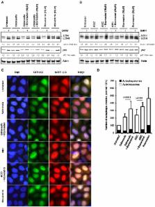
          Autophagy is a lysosomal degradation process that contributes to host immunity by eliminating invasive pathogens and the modulating inflammatory response. Several infectious and immune disorders are associated with autophagy defects, suggesting that stimulation of autophagy in these diseases should be beneficial. Here, we show that resveratrol is able to boost xenophagy, a selective form of autophagy that target invasive bacteria. We demonstrated that resveratrol promotes in vitro autophagy-dependent clearance of intracellular bacteria in intestinal epithelial cells and macrophages. These results were validated in vivo using infection in a transgenic GFP-LC3 zebrafish model. We also compared the ability of resveratrol derivatives, designed to improve the bioavailability of the parent molecule, to stimulate autophagy and to induce intracellular bacteria clearance. Together, our data demonstrate the ability of resveratrol to stimulate xenophagy, and thereby enhance the clearance of two invasive bacteria involved life-threatening diseases, Salmonella Typhimurium and Crohn's disease-associated Adherent-Invasive Escherichia coli. These findings encourage the further development of pro-autophagic nutrients to strengthen intestinal homeostasis in basal and infectious states.

          Related collections

          Most cited references56

          • Record: found
          • Abstract: found
          • Article: not found

          Loss of the autophagy protein Atg16L1 enhances endotoxin-induced IL-1beta production.

          Systems for protein degradation are essential for tight control of the inflammatory immune response. Autophagy, a bulk degradation system that delivers cytoplasmic constituents into autolysosomes, controls degradation of long-lived proteins, insoluble protein aggregates and invading microbes, and is suggested to be involved in the regulation of inflammation. However, the mechanism underlying the regulation of inflammatory response by autophagy is poorly understood. Here we show that Atg16L1 (autophagy-related 16-like 1), which is implicated in Crohn's disease, regulates endotoxin-induced inflammasome activation in mice. Atg16L1-deficiency disrupts the recruitment of the Atg12-Atg5 conjugate to the isolation membrane, resulting in a loss of microtubule-associated protein 1 light chain 3 (LC3) conjugation to phosphatidylethanolamine. Consequently, both autophagosome formation and degradation of long-lived proteins are severely impaired in Atg16L1-deficient cells. Following stimulation with lipopolysaccharide, a ligand for Toll-like receptor 4 (refs 8, 9), Atg16L1-deficient macrophages produce high amounts of the inflammatory cytokines IL-1beta and IL-18. In lipopolysaccharide-stimulated macrophages, Atg16L1-deficiency causes Toll/IL-1 receptor domain-containing adaptor inducing IFN-beta (TRIF)-dependent activation of caspase-1, leading to increased production of IL-1beta. Mice lacking Atg16L1 in haematopoietic cells are highly susceptible to dextran sulphate sodium-induced acute colitis, which is alleviated by injection of anti-IL-1beta and IL-18 antibodies, indicating the importance of Atg16L1 in the suppression of intestinal inflammation. These results demonstrate that Atg16L1 is an essential component of the autophagic machinery responsible for control of the endotoxin-induced inflammatory immune response.
            Bookmark
            • Record: found
            • Abstract: found
            • Article: not found

            Autophagy defends cells against invading group A Streptococcus.

            We found that the autophagic machinery could effectively eliminate pathogenic group A Streptococcus (GAS) within nonphagocytic cells. After escaping from endosomes into the cytoplasm, GAS became enveloped by autophagosome-like compartments and were killed upon fusion of these compartments with lysosomes. In autophagy-deficient Atg5-/- cells, GAS survived, multiplied, and were released from the cells. Thus, the autophagic machinery can act as an innate defense system against invading pathogens.
              Bookmark
              • Record: found
              • Abstract: found
              • Article: not found

              Bioavailability of resveratrol.

              This paper reviews our current understanding of the absorption, bioavailability, and metabolism of resveratrol, with an emphasis on humans. The oral absorption of resveratrol in humans is about 75% and is thought to occur mainly by transepithelial diffusion. Extensive metabolism in the intestine and liver results in an oral bioavailability considerably less than 1%. Dose escalation and repeated dose administration of resveratrol does not appear to alter this significantly. Metabolic studies, both in plasma and in urine, have revealed major metabolites to be glucuronides and sulfates of resveratrol. However, reduced dihydroresveratrol conjugates, in addition to highly polar unknown products, may account for as much as 50% of an oral resveratrol dose. Although major sites of metabolism include the intestine and liver (as expected), colonic bacterial metabolism may be more important than previously thought. Deconjugation enzymes such as β-glucuronidase and sulfatase, as well as specific tissue accumulation of resveratrol, may enhance resveratrol efficacy at target sites. Resveratrol analogs, such as methylated derivatives with improved bioavailability, may be important in future research. © 2011 New York Academy of Sciences.
                Bookmark

                Author and article information

                Contributors
                Journal
                Front Immunol
                Front Immunol
                Front. Immunol.
                Frontiers in Immunology
                Frontiers Media S.A.
                1664-3224
                14 January 2019
                2018
                : 9
                : 3149
                Affiliations
                [1] 1AgroSup Dijon, PAM UMR A 02.102, University Bourgogne Franche-Comté , Dijon, France
                [2] 2University of Bourgogne-Franche Comté , Dijon, France
                [3] 3INSERM U1231, Lipids, Nutrition Cancer , Dijon, France
                [4] 4Research Team CADIR, Cancer and Adaptative Immune Response , Dijon, France
                [5] 5UFR SVTE—UFR Sciences de la Vie, de la Terre et de l'Environnement, Université de Bourgogne Franche-Comté , Dijon, France
                [6] 6Dimacell Imaging Facility, AgroSup Dijon, University Bourgogne Franche-Comté , Dijon, France
                [7] 7AgroSup Dijon, CNRS, INRA, Centre des Sciences du Goût et de l'Alimentation, Université Bourgogne Franche-Comté , Dijon, France
                [8] 8Institut de Chimie Moléculaire de l'Université de Bourgogne (ICMUB-UMR CNRS 6302), Université of Bourgogne , Dijon, France
                Author notes

                Edited by: Zorica D. Juranic, Institute of Oncology and Radiology of Serbia, Serbia

                Reviewed by: Xi Ma, China Agricultural University, China; Zhenhuan Zhao, University of Virginia, United States

                *Correspondence: Pierre Lapaquette pierre.lapaquette@ 123456u-bourgogne.fr

                This article was submitted to Nutritional Immunology, a section of the journal Frontiers in Immunology

                †These authors share senior authorship

                Article
                10.3389/fimmu.2018.03149
                6339935
                30693000
                f2ba212f-6015-4992-9be0-ffcdb154e9e8
                Copyright © 2019 Al Azzaz, Rieu, Aires, Delmas, Chluba, Winckler, Bringer, Lamarche, Vervandier-Fasseur, Dalle, Lapaquette and Guzzo.

                This is an open-access article distributed under the terms of the Creative Commons Attribution License (CC BY). The use, distribution or reproduction in other forums is permitted, provided the original author(s) and the copyright owner(s) are credited and that the original publication in this journal is cited, in accordance with accepted academic practice. No use, distribution or reproduction is permitted which does not comply with these terms.

                History
                : 31 October 2018
                : 20 December 2018
                Page count
                Figures: 6, Tables: 1, Equations: 0, References: 76, Pages: 15, Words: 10077
                Funding
                Funded by: Association Instituts Carnot 10.13039/501100006367
                Funded by: Université de Bourgogne 10.13039/501100004618
                Categories
                Immunology
                Original Research

                Immunology
                autophagy,resveratrol,xenophagy,salmonella,aiec
                Immunology
                autophagy, resveratrol, xenophagy, salmonella, aiec

                Comments

                Comment on this article