0
views
0
recommends
+1 Recommend
0 collections
    0
    shares
      • Record: found
      • Abstract: found
      • Article: found
      Is Open Access

      Immune cell status, cardiorespiratory fitness and body composition among breast cancer survivors and healthy women: a cross sectional study

      research-article

      Read this article at

      Bookmark
          There is no author summary for this article yet. Authors can add summaries to their articles on ScienceOpen to make them more accessible to a non-specialist audience.

          Abstract

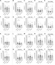
          Methods: We examined whether immune cell profiles differ between healthy women ( n = 38) and breast cancer survivors ( n = 27) within 2 years of treatment, and whether any group-differences were influenced by age, cytomegalovirus infection, cardiorespiratory fitness and body composition. Using flow cytometry, CD4+ and CD8+ T cell subsets, including naïve (NA), central memory (CM) and effector cells (EM and EMRA) were identified using CD27/CD45RA. Activation was measured by HLA-DR expression. Stem cell-like memory T cells (TSCMs) were identified using CD95/CD127. B cells, including plasmablasts, memory, immature and naïve cells were identified using CD19/CD27/CD38/CD10. Effector and regulatory Natural Killer cells were identified using CD56/CD16.

          Results: Compared to healthy women, CD4+ CM were +Δ21% higher among survivors ( p = 0.028) and CD8+ NA were −Δ25% lower ( p = 0.034). Across CD4+ and CD8+ subsets, the proportion of activated (HLA-DR+) cells was +Δ31% higher among survivors: CD4+ CM (+Δ25%), CD4+ EM (+Δ32%) and CD4+ EMRA (+Δ43%), total CD8 + (+Δ30%), CD8+ EM (+Δ30%) and CD8+ EMRA (+Δ25%) ( p < 0.046). The counts of immature B cells, NK cells and CD16+ NK effector cells were higher among survivors (+Δ100%, +Δ108% and +Δ143% respectively, p < 0.04). Subsequent analyses examined whether statistically significant differences in participant characteristics, influenced immunological differences between groups. Compared to healthy women, survivors were older (56 ± 6 y vs. 45 ± 11 y), had lower cardiorespiratory fitness ( V ˙ O 2 m a x mL kg −1 min −1: 28.8 ± 5.0 vs. 36.2 ± 8.5), lower lean mass (42.3 ± 5.0 kg vs. 48.4 ± 15.8 kg), higher body fat (36.3% ± 5.3% vs. 32.7% ± 6.4%) and higher fat mass index (FMI kg/m 2: 9.5 ± 2.2 vs. 8.1 ± 2.7) (all p < 0.033). Analysis of covariance revealed divergent moderating effects of age, CMV serostatus, cardiorespiratory fitness and body composition on the differences in immune cell profiles between groups, depending on the cell type examined. Moreover, across all participants, fat mass index was positively associated with the proportion of HLA-DR+ CD4+ EMRA and CD8+ EM/EMRA T cells (Pearson correlation: r > 0.305, p < 0.019). The association between fat mass index and HLA-DR+ CD8+ EMRA T cells withstood statistical adjustment for all variables, including age, CMV serostatus, lean mass and cardiorespiratory fitness, potentially implicating these cells as contributors to inflammatory/immune-dysfunction in overweight/obesity.

          Related collections

          Most cited references88

          • Record: found
          • Abstract: found
          • Article: not found

          A human memory T-cell subset with stem cell-like properties

          Immunological memory is thought to depend upon a stem cell-like, self-renewing population of lymphocytes capable of differentiating into effector cells in response to antigen re-exposure. Here we describe a long-lived human memory T-cell population that displays enhanced self-renewal and multipotent capacity to derive central memory, effector memory and effector T cells. These cells, specific for multiple viral and self-tumor antigens, were found within a CD45RO−, CCR7+, CD45RA+, CD62L+, CD27+, CD28+ and IL-7Rα+ T-cell compartment characteristic of naïve T cells. However, they expressed increased levels of CD95, IL-2Rβ, CXCR3, and LFA-1, and exhibited numerous functional attributes distinctive of memory cells. Compared to known memory populations, these lymphocytes displayed increased proliferative capacity, more efficiently reconstituted immunodeficient hosts and mediated superior anti-tumor responses in a humanized mouse model. The identification of a human stem cell-like memory T-cell population is of direct relevance to the design of vaccines and T-cell therapies.
            Bookmark
            • Record: found
            • Abstract: found
            • Article: found
            Is Open Access

            Anti-PD-1 and Anti-CTLA-4 Therapies in Cancer: Mechanisms of Action, Efficacy, and Limitations

            Melanoma, a skin cancer associated with high mortality rates, is highly radio- and chemotherapy resistant but can also be very immunogenic. These circumstances have led to a recent surge in research into therapies aiming to boost anti-tumor immune responses in cancer patients. Among these immunotherapies, neutralizing antibodies targeting the immune checkpoints T-lymphocyte-associated protein 4 (CTLA-4) and programmed cell death protein 1 (PD-1) are being hailed as particularly successful. These antibodies have resulted in dramatic improvements in disease outcome and are now clinically approved in many countries. However, the majority of advanced stage melanoma patients do not respond or will relapse, and the hunt for the “magic bullet” to treat the disease continues. This review examines the mechanisms of action and the limitations of anti-PD-1/PD-L1 and anti-CTLA-4 antibodies which are the two types of checkpoint inhibitors currently available to patients and further explores the future avenues of their use in melanoma and other cancers.
              Bookmark
              • Record: found
              • Abstract: found
              • Article: not found

              The biology of human natural killer-cell subsets.

              Human natural killer (NK) cells comprise approximately 15% of all circulating lymphocytes. Owing to their early production of cytokines and chemokines, and ability to lyse target cells without prior sensitization, NK cells are crucial components of the innate immune system. Human NK cells can be divided into two subsets based on their cell-surface density of CD56--CD56(bright) and CD56(dim)--each with distinct phenotypic properties. Now, there is ample evidence to suggest that these NK-cell subsets have unique functional attributes and, therefore, distinct roles in the human immune response. The CD56(dim) NK-cell subset is more naturally cytotoxic and expresses higher levels of Ig-like NK receptors and FCgamma receptor III (CD16) than the CD56(bright) NK-cell subset. By contrast, the CD56(bright) subset has the capacity to produce abundant cytokines following activation of monocytes, but has low natural cytotoxicity and is CD16(dim) or CD16(-). In addition, we will discuss other cell-surface receptors expressed differentially by human NK-cell subsets and the distinct functional properties of these subsets.
                Bookmark

                Author and article information

                Contributors
                Journal
                Front Physiol
                Front Physiol
                Front. Physiol.
                Frontiers in Physiology
                Frontiers Media S.A.
                1664-042X
                01 June 2023
                2023
                : 14
                : 1107070
                Affiliations
                [1] 1 Department for Health , University of Bath , Bath, United Kingdom
                [2] 2 Department for Oncology and Haematology , Royal United Hospitals Bath NHS Trust , Bath, United Kingdom
                [3] 3 Velindre Cancer Centre and Cardiff University , Cardiff, United Kingdom
                [4] 4 School of Sport, Exercise and Rehabilitation Sciences , College of Life and Environmental Sciences , University of Birmingham , Birmingham, United Kingdom
                Author notes

                Edited by: Christian Puta, Friedrich Schiller University Jena, Germany

                Reviewed by: Emily C. LaVoy, University of Houston, United States

                Wilhelm Bloch, German Sport University Cologne, Germany

                *Correspondence: James E. Turner, j.e.turner.2@ 123456bham.ac.uk
                Article
                1107070
                10.3389/fphys.2023.1107070
                10267418
                37324393
                f41755a1-72a1-4615-a77c-91b7cd2e32bc
                Copyright © 2023 Arana Echarri, Struszczak, Beresford, Campbell, Jones, Thompson and Turner.

                This is an open-access article distributed under the terms of the Creative Commons Attribution License (CC BY). The use, distribution or reproduction in other forums is permitted, provided the original author(s) and the copyright owner(s) are credited and that the original publication in this journal is cited, in accordance with accepted academic practice. No use, distribution or reproduction is permitted which does not comply with these terms.

                History
                : 24 November 2022
                : 19 May 2023
                Funding
                This work was supported in part by grant MR/N0137941/1 for the GW4 BIOMED MRC DTP, awarded to the Universities of Bath, Bristol, Cardiff and Exeter from the Medical Research Council (MRC)/UKRI.
                Categories
                Physiology
                Original Research
                Custom metadata
                Exercise Physiology

                Anatomy & Physiology
                breast cancer treatment,immune profiles,leukocytes,lymphocytes,lifestyle,exercise,cardiorespiratory fitness,body composition

                Comments

                Comment on this article