Inviting an author to review:
Find an author and click ‘Invite to review selected article’ near their name.
Search for authorsSearch for similar articles
26
views
0
recommends
+1 Recommend
0 collections
    0
    shares
      • Record: found
      • Abstract: found
      • Article: found
      Is Open Access

      Thyroxine Differentially Modulates the Peripheral Clock: Lessons from the Human Hair Follicle

      research-article

      Read this article at

      Bookmark
          There is no author summary for this article yet. Authors can add summaries to their articles on ScienceOpen to make them more accessible to a non-specialist audience.

          Abstract

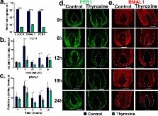
          The human hair follicle (HF) exhibits peripheral clock activity, with knock-down of clock genes ( BMAL1 and PER1) prolonging active hair growth (anagen) and increasing pigmentation. Similarly, thyroid hormones prolong anagen and stimulate pigmentation in cultured human HFs. In addition they are recognized as key regulators of the central clock that controls circadian rhythmicity. Therefore, we asked whether thyroxine (T4) also influences peripheral clock activity in the human HF. Over 24 hours we found a significant reduction in protein levels of BMAL1 and PER1, with their transcript levels also decreasing significantly. Furthermore, while all clock genes maintained their rhythmicity in both the control and T4 treated HFs, there was a significant reduction in the amplitude of BMAL1 and PER1 in T4 (100 nM) treated HFs. Accompanying this, cell-cycle progression marker Cyclin D1 was also assessed appearing to show an induced circadian rhythmicity by T4 however, this was not significant. Contrary to short term cultures, after 6 days, transcript and/or protein levels of all core clock genes (BMAL1, PER1, clock, CRY1, CRY2) were up-regulated in T4 treated HFs. BMAL1 and PER1 mRNA was also up-regulated in the HF bulge, the location of HF epithelial stem cells. Together this provides the first direct evidence that T4 modulates the expression of the peripheral molecular clock. Thus, patients with thyroid dysfunction may also show a disordered peripheral clock, which raises the possibility that short term, pulsatile treatment with T4 might permit one to modulate circadian activity in peripheral tissues as a target to treat clock-related disease.

          Related collections

          Most cited references50

          • Record: found
          • Abstract: found
          • Article: not found

          Molecular architecture of the mammalian circadian clock.

          Circadian clocks coordinate physiology and behavior with the 24h solar day to provide temporal homeostasis with the external environment. The molecular clocks that drive these intrinsic rhythmic changes are based on interlocked transcription/translation feedback loops that integrate with diverse environmental and metabolic stimuli to generate internal 24h timing. In this review we highlight recent advances in our understanding of the core molecular clock and how it utilizes diverse transcriptional and post-transcriptional mechanisms to impart temporal control onto mammalian physiology. Understanding the way in which biological rhythms are generated throughout the body may provide avenues for temporally directed therapeutics to improve health and prevent disease. Copyright © 2013 Elsevier Ltd. All rights reserved.
            Bookmark
            • Record: found
            • Abstract: found
            • Article: not found

            Resetting of circadian time in peripheral tissues by glucocorticoid signaling.

            In mammals, circadian oscillators reside not only in the suprachiasmatic nucleus of the brain, which harbors the central pacemaker, but also in most peripheral tissues. Here, we show that the glucocorticoid hormone analog dexamethasone induces circadian gene expression in cultured rat-1 fibroblasts and transiently changes the phase of circadian gene expression in liver, kidney, and heart. However, dexamethasone does not affect cyclic gene expression in neurons of the suprachiasmatic nucleus. This enabled us to establish an apparent phase-shift response curve specifically for peripheral clocks in intact animals. In contrast to the central clock, circadian oscillators in peripheral tissues appear to remain responsive to phase resetting throughout the day.
              Bookmark
              • Record: found
              • Abstract: found
              • Article: not found

              Early aging and age-related pathologies in mice deficient in BMAL1, the core componentof the circadian clock.

              Mice deficient in the circadian transcription factor BMAL1 (brain and muscle ARNT-like protein) have impaired circadian behavior and demonstrate loss of rhythmicity in the expression of target genes. Here we report that Bmal1(-/-) mice have reduced lifespans and display various symptoms of premature aging including sarcopenia, cataracts, less subcutaneous fat, organ shrinkage, and others. The early aging phenotype correlates with increased levels of reactive oxygen species in some tissues of the Bmal1(-/- )animals. These findings, together with data on CLOCK/BMAL1-dependent control of stress responses, may provide a mechanistic explanation for the early onset of age-related pathologies in the absence of BMAL1.
                Bookmark

                Author and article information

                Contributors
                Role: Academic Editor
                Journal
                PLoS One
                PLoS ONE
                plos
                plosone
                PLoS ONE
                Public Library of Science (San Francisco, CA USA )
                1932-6203
                30 March 2015
                2015
                : 10
                : 3
                : e0121878
                Affiliations
                [1 ]The Dermatology Centre, Institute of Inflammation and Repair, University of Manchester, Manchester, United Kingdom
                [2 ]Doctoral Training Centre in Integrative Systems Biology, Manchester Interdisciplinary Bio centre, University of Manchester, Manchester, United Kingdom
                [3 ]The Farjo Hair Institute, Manchester, United Kingdom
                [4 ]Department of Dermatology, University of Muenster, Muenster, Germany
                University of Texas Southwestern Medical Center, UNITED STATES
                Author notes

                Competing Interests: The authors have declared that no competing interests exist.

                Conceived and designed the experiments: JH RP. Performed the experiments: JH. Analyzed the data: JH. Contributed reagents/materials/analysis tools: NF BF IH. Wrote the paper: JH IH RP.

                Article
                PONE-D-14-34544
                10.1371/journal.pone.0121878
                4379003
                25822259
                f766a7da-dbd3-47f1-b0a8-eb6be3549e1a
                Copyright @ 2015

                This is an open access article distributed under the terms of the Creative Commons Attribution License, which permits unrestricted use, distribution, and reproduction in any medium, provided the original author and source are credited

                History
                : 1 August 2014
                : 18 February 2015
                Page count
                Figures: 4, Tables: 0, Pages: 15
                Funding
                This work was funded by a grant given to JH to complete his PhD studies from the Biology and biotechnology research council (BBSRC), however the authors received no specific funding for this work. Human tissue was stored in a registered biobank following human tissue act guidelines and stored with ethical and institutional approval from the University of Manchester.
                Categories
                Research Article
                Custom metadata
                All data relevant to this investigation can be found in the manuscript and supplementary files.

                Uncategorized
                Uncategorized

                Comments

                Comment on this article