30
views
0
recommends
+1 Recommend
0 collections
    0
    shares
      • Record: found
      • Abstract: found
      • Article: found
      Is Open Access

      Cell Senescence: A Nonnegligible Cell State under Survival Stress in Pathology of Intervertebral Disc Degeneration

      review-article

      Read this article at

      Bookmark
          There is no author summary for this article yet. Authors can add summaries to their articles on ScienceOpen to make them more accessible to a non-specialist audience.

          Abstract

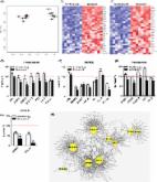
          The intervertebral disc degeneration (IDD) with increasing aging mainly manifests as low back pain (LBP) accompanied with a loss of physical ability. These pathological processes can be preliminarily interpreted as a series of changes at cellular level. In addition to cell death, disc cells enter into the stagnation with dysfunction and deteriorate tissue microenvironment in degenerative discs, which is recognized as cell senescence. During aging, many intrinsic and extrinsic factors have been proved to have strong connections with these cellular senescence phenomena. Growing evidences of these connections require us to gather up critical cues from potential risk factors to pathogenesis and relative interventions for retarding cell senescence and attenuating degenerative changes. In this paper, we try to clarify another important cell state apart from cell death in IDD and discuss senescence-associated changes in cells and extracellular microenvironment. Then, we emphasize the role of oxidative stress and epigenomic perturbations in linking risk factors to cell senescence in the onset of IDD. Further, we summarize the current interventions targeting senescent cells that may exert the benefits of antidegeneration in IDD.

          Related collections

          Most cited references92

          • Record: found
          • Abstract: found
          • Article: not found

          Cellular senescence in aging and age-related disease: from mechanisms to therapy.

          Cellular senescence, a process that imposes permanent proliferative arrest on cells in response to various stressors, has emerged as a potentially important contributor to aging and age-related disease, and it is an attractive target for therapeutic exploitation. A wealth of information about senescence in cultured cells has been acquired over the past half century; however, senescence in living organisms is poorly understood, largely because of technical limitations relating to the identification and characterization of senescent cells in tissues and organs. Furthermore, newly recognized beneficial signaling functions of senescence suggest that indiscriminately targeting senescent cells or modulating their secretome for anti-aging therapy may have negative consequences. Here we discuss current progress and challenges in understanding the stressors that induce senescence in vivo, the cell types that are prone to senesce, and the autocrine and paracrine properties of senescent cells in the contexts of aging and age-related diseases as well as disease therapy.
            Bookmark
            • Record: found
            • Abstract: found
            • Article: found
            Is Open Access

            The Achilles’ heel of senescent cells: from transcriptome to senolytic drugs

            The healthspan of mice is enhanced by killing senescent cells using a transgenic suicide gene. Achieving the same using small molecules would have a tremendous impact on quality of life and the burden of age-related chronic diseases. Here, we describe the rationale for identification and validation of a new class of drugs termed senolytics, which selectively kill senescent cells. By transcript analysis, we discovered increased expression of pro-survival networks in senescent cells, consistent with their established resistance to apoptosis. Using siRNA to silence expression of key nodes of this network, including ephrins (EFNB1 or 3), PI3Kδ, p21, BCL-xL, or plasminogen-activated inhibitor-2, killed senescent cells, but not proliferating or quiescent, differentiated cells. Drugs targeting these same factors selectively killed senescent cells. Dasatinib eliminated senescent human fat cell progenitors, while quercetin was more effective against senescent human endothelial cells and mouse BM-MSCs. The combination of dasatinib and quercetin was effective in eliminating senescent MEFs. In vivo, this combination reduced senescent cell burden in chronologically aged, radiation-exposed, and progeroid Ercc1 −/Δ mice. In old mice, cardiac function and carotid vascular reactivity were improved 5 days after a single dose. Following irradiation of one limb in mice, a single dose led to improved exercise capacity for at least 7 months following drug treatment. Periodic drug administration extended healthspan in Ercc1 −/Δ mice, delaying age-related symptoms and pathology, osteoporosis, and loss of intervertebral disk proteoglycans. These results demonstrate the feasibility of selectively ablating senescent cells and the efficacy of senolytics for alleviating symptoms of frailty and extending healthspan.
              Bookmark
              • Record: found
              • Abstract: found
              • Article: not found

              Senescence and tumour clearance is triggered by p53 restoration in murine liver carcinomas.

              Although cancer arises from a combination of mutations in oncogenes and tumour suppressor genes, the extent to which tumour suppressor gene loss is required for maintaining established tumours is poorly understood. p53 is an important tumour suppressor that acts to restrict proliferation in response to DNA damage or deregulation of mitogenic oncogenes, by leading to the induction of various cell cycle checkpoints, apoptosis or cellular senescence. Consequently, p53 mutations increase cell proliferation and survival, and in some settings promote genomic instability and resistance to certain chemotherapies. To determine the consequences of reactivating the p53 pathway in tumours, we used RNA interference (RNAi) to conditionally regulate endogenous p53 expression in a mosaic mouse model of liver carcinoma. We show that even brief reactivation of endogenous p53 in p53-deficient tumours can produce complete tumour regressions. The primary response to p53 was not apoptosis, but instead involved the induction of a cellular senescence program that was associated with differentiation and the upregulation of inflammatory cytokines. This program, although producing only cell cycle arrest in vitro, also triggered an innate immune response that targeted the tumour cells in vivo, thereby contributing to tumour clearance. Our study indicates that p53 loss can be required for the maintenance of aggressive carcinomas, and illustrates how the cellular senescence program can act together with the innate immune system to potently limit tumour growth.
                Bookmark

                Author and article information

                Contributors
                Journal
                Oxid Med Cell Longev
                Oxid Med Cell Longev
                OMCL
                Oxidative Medicine and Cellular Longevity
                Hindawi
                1942-0900
                1942-0994
                2020
                31 August 2020
                : 2020
                : 9503562
                Affiliations
                1Department of Orthopedics Surgery, The Second Affiliated Hospital, Zhejiang University School of Medicine, 310009 Hangzhou, Zhejiang, China
                2Zhejiang Key Laboratory of Bone and Joint Precision and Department of Orthopedics Research Institute of Zhejiang University, Hangzhou, Zhejiang 310009, China
                Author notes

                Academic Editor: Wenyuan Ding

                Author information
                https://orcid.org/0000-0002-2402-781X
                https://orcid.org/0000-0002-4983-6461
                https://orcid.org/0000-0002-2626-0201
                Article
                10.1155/2020/9503562
                7479476
                32934764
                fa2164a2-743e-4963-bd00-f307bc5301a4
                Copyright © 2020 Yuang Zhang et al.

                This is an open access article distributed under the Creative Commons Attribution License, which permits unrestricted use, distribution, and reproduction in any medium, provided the original work is properly cited.

                History
                : 3 July 2020
                : 1 August 2020
                : 10 August 2020
                Funding
                Funded by: National Natural Science Foundation of China
                Award ID: 81772379
                Award ID: 81902238
                Award ID: 81972096
                Funded by: Natural Science Foundation of Zhejiang Province
                Award ID: LY19H060005
                Award ID: LQ18H060003
                Funded by: Scientific Research Fund of Zhejiang Provincial Education Department
                Award ID: Y201941491
                Award ID: Y201941476
                Funded by: China Postdoctoral Science Foundation
                Award ID: 2017M612011
                Funded by: Medical Science and Technology Project of Zhejiang Province of China
                Award ID: 2020385155
                Categories
                Review Article

                Molecular medicine
                Molecular medicine

                Comments

                Comment on this article