Inviting an author to review:
Find an author and click ‘Invite to review selected article’ near their name.
Search for authorsSearch for similar articles
5
views
0
recommends
+1 Recommend
0 collections
    0
    shares
      • Record: found
      • Abstract: found
      • Article: found
      Is Open Access

      Acetaminophen impairs ferroptosis in the hippocampus of septic mice by regulating glutathione peroxidase 4 and ferroptosis suppressor protein 1 pathways

      research-article

      Read this article at

      Bookmark
          There is no author summary for this article yet. Authors can add summaries to their articles on ScienceOpen to make them more accessible to a non-specialist audience.

          Abstract

          Background

          Neuronal ferroptosis is a major cause of cognitive impairment and mortality in patients with sepsis‐associated encephalopathy (SAE). A low dose of acetaminophen (APAP) in septic mice can prevent ferroptosis in the hippocampal tissue; however, the underlying mechanism is unknown. This study aimed to investigate the mechanism by which APAP reduces ferroptosis in the hippocampal tissues of septic mice.

          Methods

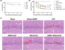
          A mouse model of SAE was established, and the ferroptosis pathway inhibitors RSL3 and iFSP1+RSL3 were used in addition to APAP for the interventions, respectively. The 7‐day survival rate of the mice was recorded, and cognitive function was examined using the Morris water maze test. Hematoxylin and eosin staining was performed to observe hippocampal tissue damage. Hippocampal iron and malondialdehyde (MDA) were measured using chemical colorimetric methods. Immunofluorescence was used to detect the reactive oxygen species (ROS) content in hippocampal tissues.

          Results

          RSL3 reversed the efficacy of APAP on improving cognitive dysfunction in septic mice but did not obviously reverse the survival rate of mice enhanced by APAP. RSL3 aggravated APAP‐induced hippocampal tissue damage in mice attenuated by APAP. RSL3 inhibited glutathione peroxidase 4 (GPX4) expression and increased ferroptosis suppressor protein 1 (FSP1) and 4‐hydroxy‐2‐nonenal (4‐HNE) expression. RSL3 also reversed the effects of APAP in reducing iron, MDA, and ROS levels in the hippocampal tissues of septic mice. iFSP1+RSL3 further reversed the effect of APAP on ameliorating cognitive dysfunction in septic mice and successfully reversed the survival rate of mice enhanced by APAP. iFSP1+RSL3 aggravated APAP‐induced cerebral hippocampal damage. iFSP1+RSL3 inhibited both GPX4 and FSP1, further reversing the effect of APAP on the reduction in iron, 4‐HNE, ROS, and MDA levels in the cerebral hippocampus of mice with sepsis.

          Conclusion

          These data suggest that APAP inhibits ferroptosis in the cerebral hippocampus of septic mice through the GPX4 and FSP1 pathways.

          Abstract

          Neuronal ferroptosis is a major cause of cognitive impairment and mortality in sepsis‐associated encephalopathy (SAE). In septic mice, a little dosage of acetaminophen (APAP) can prevent hippocampus tissue ferroptosis, however the mechanism is unknown. The goal of this study was to investigate the mechanism through which APAP reduces ferroptosis in septic mice hippocampus tissue.

          Related collections

          Most cited references19

          • Record: found
          • Abstract: found
          • Article: not found

          Ferroptosis: A Regulated Cell Death Nexus Linking Metabolism, Redox Biology, and Disease

          Ferroptosis is a form of regulated cell death characterized by the iron-dependent accumulation of lipid hydroperoxides to lethal levels. Emerging evidence suggests that ferroptosis represents an ancient vulnerability caused by the incorporation of polyunsaturated fatty acids into cellular membranes, and cells have developed complex systems that exploit and defend against this vulnerability in different contexts. The sensitivity to ferroptosis is tightly linked to numerous biological processes, including amino acid, iron, and polyunsaturated fatty acid metabolism, and the biosynthesis of glutathione, phospholipids, NADPH, and coenzyme Q10. Ferroptosis has been implicated in the pathological cell death associated with degenerative diseases (i.e., Alzheimer's, Huntington's, and Parkinson's diseases), carcinogenesis, stroke, intracerebral hemorrhage, traumatic brain injury, ischemia-reperfusion injury, and kidney degeneration in mammals and is also implicated in heat stress in plants. Ferroptosis may also have a tumor-suppressor function that could be harnessed for cancer therapy. This Primer reviews the mechanisms underlying ferroptosis, highlights connections to other areas of biology and medicine, and recommends tools and guidelines for studying this emerging form of regulated cell death.
            Bookmark
            • Record: found
            • Abstract: found
            • Article: not found

            The CoQ oxidoreductase FSP1 acts in parallel to GPX4 to inhibit ferroptosis

            Ferroptosis is a form of regulated cell death that is caused by the iron-dependent peroxidation of lipids 1,2 . The glutathione-dependent lipid hydroperoxidase glutathione peroxidase 4 (GPX4) prevents ferroptosis by converting lipid hydroperoxides into non-toxic lipid alcohols 3,4 . Ferroptosis has been implicated in the cell death that underlies several degenerative conditions 2 , and induction of ferroptosis by inhibition of GPX4 has emerged as a therapeutic strategy to trigger cancer cell death 5 . However, sensitivity to GPX4 inhibitors varies greatly across cancer cell lines 6 , suggesting that additional factors govern resistance to ferroptosis. Here, employing a synthetic lethal CRISPR/Cas9 screen, we identify ferroptosis suppressor protein 1 (FSP1) (previously known as apoptosis-inducing factor mitochondrial 2 (AIFM2)) as a potent ferroptosis resistance factor. Our data indicate that myristoylation recruits FSP1 to the plasma membrane where it functions as an oxidoreductase that reduces coenzyme Q10 (CoQ), generating a lipophilic radical-trapping antioxidant (RTA) that halts the propagation of lipid peroxides. We further find that FSP1 expression positively correlates with ferroptosis resistance across hundreds of cancer cell lines, and that FSP1 mediates resistance to ferroptosis in lung cancer cells in culture and in mouse tumor xenografts. Thus, our data identify FSP1 as a key component of a non-mitochondrial CoQ antioxidant system that acts in parallel to the canonical glutathione-based GPX4 pathway. These findings define a new ferroptosis suppression pathway and indicate that pharmacological inhibition of FSP1 may provide an effective strategy to sensitize cancer cells to ferroptosis-inducing chemotherapeutics.
              Bookmark
              • Record: found
              • Abstract: not found
              • Article: not found

              FSP1 is a glutathione-independent ferroptosis suppressor

                Bookmark

                Author and article information

                Contributors
                yyu77@tmu.edu.cn
                yuyonghao@126.com
                Journal
                Brain Behav
                Brain Behav
                10.1002/(ISSN)2157-9032
                BRB3
                Brain and Behavior
                John Wiley and Sons Inc. (Hoboken )
                2162-3279
                13 July 2023
                August 2023
                : 13
                : 8 ( doiID: 10.1002/brb3.v13.8 )
                : e3145
                Affiliations
                [ 1 ] Department of Anesthesiology Characteristic Medical Center of Chinese People's Armed Police Force (PAP) Tianjin China
                [ 2 ] Department of Anesthesiology Tianjin Medical University General Hospital Tianjin China
                [ 3 ] Tianjin Institute of Anesthesiology Tianjin China
                [ 4 ] Department of Anesthesiology Tianjin Children's Hospital Tianjin China
                Author notes
                [*] [* ] Correspondence

                Yang Yu and Yonghao Yu, Department of Anesthesiology, Tianjin Medical University General Hospital, No. 154 Anshan Rd, Heping District, Tianjin 300052, China.

                Email: yyu77@ 123456tmu.edu.cn and yuyonghao@ 123456126.com

                [#]

                Jing Chu, Hong Li, and Zhihao Yuan contributed equally to the work.

                Author information
                https://orcid.org/0000-0002-8303-8690
                Article
                BRB33145
                10.1002/brb3.3145
                10454284
                37443407
                fe39b73a-1f49-4a9e-af01-c9d9cbfa45af
                © 2023 The Authors. Brain and Behavior published by Wiley Periodicals LLC.

                This is an open access article under the terms of the http://creativecommons.org/licenses/by/4.0/ License, which permits use, distribution and reproduction in any medium, provided the original work is properly cited.

                History
                : 02 June 2023
                : 30 January 2023
                : 20 June 2023
                Page count
                Figures: 10, Tables: 0, Pages: 13, Words: 7085
                Funding
                Funded by: National Natural Science Foundation of China , doi 10.13039/501100001809;
                Award ID: 82001149
                Award ID: 82072150
                Categories
                Original Article
                Original Articles
                Custom metadata
                2.0
                August 2023
                Converter:WILEY_ML3GV2_TO_JATSPMC version:6.3.3 mode:remove_FC converted:25.08.2023

                Neurosciences
                acetaminophen,ferroptosis,fsp1,gpx4,sepsis‐associated encephalopathy
                Neurosciences
                acetaminophen, ferroptosis, fsp1, gpx4, sepsis‐associated encephalopathy

                Comments

                Comment on this article