21
views
0
recommends
+1 Recommend
0 collections
    0
    shares
      • Record: found
      • Abstract: found
      • Article: found
      Is Open Access

      Complement-dependent outer membrane perturbation sensitizes Gram-negative bacteria to Gram-positive specific antibiotics

      research-article

      Read this article at

      Bookmark
          There is no author summary for this article yet. Authors can add summaries to their articles on ScienceOpen to make them more accessible to a non-specialist audience.

          Abstract

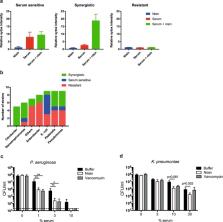
          Gram-negative bacteria are refractory to the action of many antibiotics due to their impermeable outer membrane. An important player of the immune system is the complement system, a protein network in serum that directly kills Gram-negative bacteria through pore-formation by the Membrane Attack Complexes (MAC). We here show that the MAC rapidly perforates the outer membrane but that inner membrane damage, which is essential for killing, is relatively slow. Importantly, we demonstrate that MAC-induced outer membrane damage sensitizes Gram-negative bacteria to otherwise ineffective, Gram-positive-specific, antimicrobials. Synergy between serum and nisin was observed for 22 out of 53 tested Gram-negative clinical isolates and for multi-drug resistant (MDR) blood isolates. The in vivo relevance of this process is further highlighted by the fact that blood sensitizes a MDR K. pneumoniae strain to vancomycin. Altogether, these data imply that antibiotics that are considered ineffective to treat infections with Gram-negatives may have different functional outcomes in patients, due to the presence of the complement system.

          Related collections

          Most cited references33

          • Record: found
          • Abstract: found
          • Article: not found

          Outer membrane permeability and antibiotic resistance.

          To date most antibiotics are targeted at intracellular processes, and must be able to penetrate the bacterial cell envelope. In particular, the outer membrane of gram-negative bacteria provides a formidable barrier that must be overcome. There are essentially two pathways that antibiotics can take through the outer membrane: a lipid-mediated pathway for hydrophobic antibiotics, and general diffusion porins for hydrophilic antibiotics. The lipid and protein compositions of the outer membrane have a strong impact on the sensitivity of bacteria to many types of antibiotics, and drug resistance involving modifications of these macromolecules is common. This review will describe the molecular mechanisms for permeation of antibiotics through the outer membrane, and the strategies that bacteria have deployed to resist antibiotics by modifications of these pathways.
            Bookmark
            • Record: found
            • Abstract: found
            • Article: not found

            Specific binding of nisin to the peptidoglycan precursor lipid II combines pore formation and inhibition of cell wall biosynthesis for potent antibiotic activity.

            Unlike numerous pore-forming amphiphilic peptide antibiotics, the lantibiotic nisin is active in nanomolar concentrations, which results from its ability to use the lipid-bound cell wall precursor lipid II as a docking molecule for subsequent pore formation. Here we use genetically engineered nisin variants to identify the structural requirements for the interaction of the peptide with lipid II. Mutations affecting the conformation of the N-terminal part of nisin comprising rings A through C, e.g. [S3T]nisin, led to reduced binding and increased the peptide concentration necessary for pore formation. The binding constant for the S3T mutant was 0.043 x 10(7) m(-1) compared with 2 x 10(7) m(-1) for the wild-type peptide, and the minimum concentration for pore formation increased from the 1 nm to the 50 nm range. In contrast, peptides mutated in the flexible hinge region, e.g. [DeltaN20/DeltaM21]nisin, were completely inactive in the pore formation assay, but were reduced to some extent in their in vivo activity. We found the remaining in vivo activity to result from the unaltered capacity of the mutated peptide to bind to lipid II and thus to inhibit its incorporation into the peptidoglycan network. Therefore, through interaction with the membrane-bound cell wall precursor lipid II, nisin inhibits peptidoglycan synthesis and forms highly specific pores. The combination of two killing mechanisms in one molecule potentiates antibiotic activity and results in nanomolar MIC values, a strategy that may well be worth considering for the construction of novel antibiotics.
              Bookmark
              • Record: found
              • Abstract: found
              • Article: not found

              Lipid II as a target for antibiotics.

              Lipid II is a membrane-anchored cell-wall precursor that is essential for bacterial cell-wall biosynthesis. The effectiveness of targeting Lipid II as an antibacterial strategy is highlighted by the fact that it is the target for at least four different classes of antibiotic, including the clinically important glycopeptide antibiotic vancomycin. However, the growing problem of bacterial resistance to many current drugs, including vancomycin, has led to increasing interest in the therapeutic potential of other classes of compound that target Lipid II. Here, we review progress in understanding of the antibacterial activities of these compounds, which include lantibiotics, mannopeptimycins and ramoplanin, and consider factors that will be important in exploiting their potential as new treatments for bacterial infections.
                Bookmark

                Author and article information

                Contributors
                b.w.bardoel-2@umcutrecht.nl
                Journal
                Sci Rep
                Sci Rep
                Scientific Reports
                Nature Publishing Group UK (London )
                2045-2322
                28 February 2019
                28 February 2019
                2019
                : 9
                : 3074
                Affiliations
                [1 ]Medical Microbiology, University Medical Center Utrecht, Utrecht University, Utrecht, Netherlands
                [2 ]ISNI 0000 0001 2312 1970, GRID grid.5132.5, Institute of Biology Leiden, , Leiden University, ; Leiden, Netherlands
                [3 ]ISNI 0000000120346234, GRID grid.5477.1, Department of Biochemistry and Cell Biology, , Utrecht University, ; Utrecht, Netherlands
                Author information
                http://orcid.org/0000-0001-6450-277X
                Article
                38577
                10.1038/s41598-019-38577-9
                6395757
                30816122
                fe6fd665-5a64-4454-8af4-cddede85225c
                © The Author(s) 2019

                Open Access This article is licensed under a Creative Commons Attribution 4.0 International License, which permits use, sharing, adaptation, distribution and reproduction in any medium or format, as long as you give appropriate credit to the original author(s) and the source, provide a link to the Creative Commons license, and indicate if changes were made. The images or other third party material in this article are included in the article’s Creative Commons license, unless indicated otherwise in a credit line to the material. If material is not included in the article’s Creative Commons license and your intended use is not permitted by statutory regulation or exceeds the permitted use, you will need to obtain permission directly from the copyright holder. To view a copy of this license, visit http://creativecommons.org/licenses/by/4.0/.

                History
                : 19 October 2018
                : 2 January 2019
                Funding
                Funded by: FundRef https://doi.org/10.13039/501100001826, ZonMw (Netherlands Organisation for Health Research and Development);
                Award ID: 541001001
                Award ID: 541001001
                Award Recipient :
                Categories
                Article
                Custom metadata
                © The Author(s) 2019

                Uncategorized
                Uncategorized

                Comments

                Comment on this article