- Record: found
- Abstract: found
- Article: found
Reducing scan time in 177Lu planar scintigraphy using convolutional neural network: A Monte Carlo simulation study
Read this article at
Abstract
Purpose
The aim of this study was to reduce scan time in 177Lu planar scintigraphy through the use of convolutional neural network (CNN) to facilitate personalized dosimetry for 177Lu‐based peptide receptor radionuclide therapy.
Methods
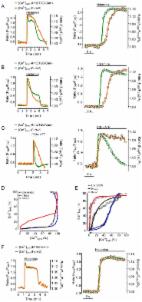
The CNN model used in this work was based on DenseNet, and the training and testing datasets were generated from Monte Carlo simulation. The CNN input images (IMG input) consisted of 177Lu planar scintigraphy that contained 10–90% of the total photon counts, while the corresponding full‐count images (IMG 100%) were used as the CNN label images. Two‐sample t‐test was conducted to compare the difference in pixel intensities within region of interest between IMG 100% and CNN output images (IMG output).
Results
No difference was found in IMG output for rods with diameters ranging from 13 to 33 mm in the Derenzo phantom with a target‐to‐background ratio of 20:1, while statistically significant differences were found in IMG output for the 10‐mm diameter rods when IMG input containing 10% to 60% of the total photon counts were denoised. Statistically significant differences were found in IMG output for both right and left kidneys in the NCAT phantom when IMG input containing 10% of the total photon counts were denoised. No statistically significant differences were found in IMG output for any other source organs in the NCAT phantom.
Related collections
Most cited references28
- Record: found
- Abstract: not found
- Article: not found
A Threshold Selection Method from Gray-Level Histograms
- Record: found
- Abstract: found
- Article: not found
GATE: a simulation toolkit for PET and SPECT.
- Record: found
- Abstract: found
- Article: not found