8
views
0
recommends
+1 Recommend
0 collections
    0
    shares
      • Record: found
      • Abstract: found
      • Article: found
      Is Open Access

      Depressed neuromuscular transmission causes weakness in mice lacking BK potassium channels

      research-article

      Read this article at

      Bookmark
          There is no author summary for this article yet. Authors can add summaries to their articles on ScienceOpen to make them more accessible to a non-specialist audience.

          Abstract

          Mice lacking BK potassium channels have weakness with stimulation of peripheral nerve, but not muscle, which is caused by a defect in neuromuscular transmission. Prolonging the motor neuron action potential fully normalizes in vivo strength.

          Abstract

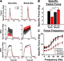
          Mice lacking functional large-conductance voltage- and Ca 2+-activated K + channels (BK channels) are viable but have motor deficits including ataxia and weakness. The cause of weakness is unknown. In this study, we discovered, in vivo, that skeletal muscle in mice lacking BK channels (BK −/−) was weak in response to nerve stimulation but not to direct muscle stimulation, suggesting a failure of neuromuscular transmission. Voltage-clamp studies of the BK −/− neuromuscular junction (NMJ) revealed a reduction in evoked endplate current amplitude and the frequency of spontaneous vesicle release compared with WT littermates. Responses to 50-Hz stimulation indicated a reduced probability of vesicle release in BK −/− mice, suggestive of lower presynaptic Ca 2+ entry. Pharmacological block of BK channels in WT NMJs did not affect NMJ function, surprisingly suggesting that the reduced vesicle release in BK −/− NMJs was not due to loss of BK channel–mediated K + current. Possible explanations for our data include an effect of BK channels on development of the NMJ, a role for BK channels in regulating presynaptic Ca 2+ current or the effectiveness of Ca 2+ in triggering release. Consistent with reduced Ca 2+ entry or effectiveness of Ca 2+ in triggering release, use of 3,4-diaminopyridine to widen action potentials normalized evoked release in BK −/− mice to WT levels. Intraperitoneal application of 3,4-diaminopyridine fully restored in vivo nerve-stimulated muscle force in BK −/− mice. Our work demonstrates that mice lacking BK channels have weakness due to a defect in vesicle release at the NMJ.

          Related collections

          Most cited references53

          • Record: found
          • Abstract: found
          • Article: not found

          Analysis of relative gene expression data using real-time quantitative PCR and the 2(-Delta Delta C(T)) Method.

          The two most commonly used methods to analyze data from real-time, quantitative PCR experiments are absolute quantification and relative quantification. Absolute quantification determines the input copy number, usually by relating the PCR signal to a standard curve. Relative quantification relates the PCR signal of the target transcript in a treatment group to that of another sample such as an untreated control. The 2(-Delta Delta C(T)) method is a convenient way to analyze the relative changes in gene expression from real-time quantitative PCR experiments. The purpose of this report is to present the derivation, assumptions, and applications of the 2(-Delta Delta C(T)) method. In addition, we present the derivation and applications of two variations of the 2(-Delta Delta C(T)) method that may be useful in the analysis of real-time, quantitative PCR data. Copyright 2001 Elsevier Science (USA).
            Bookmark
            • Record: found
            • Abstract: found
            • Article: not found

            Short-term synaptic plasticity.

            Synaptic transmission is a dynamic process. Postsynaptic responses wax and wane as presynaptic activity evolves. This prominent characteristic of chemical synaptic transmission is a crucial determinant of the response properties of synapses and, in turn, of the stimulus properties selected by neural networks and of the patterns of activity generated by those networks. This review focuses on synaptic changes that result from prior activity in the synapse under study, and is restricted to short-term effects that last for at most a few minutes. Forms of synaptic enhancement, such as facilitation, augmentation, and post-tetanic potentiation, are usually attributed to effects of a residual elevation in presynaptic [Ca(2+)]i, acting on one or more molecular targets that appear to be distinct from the secretory trigger responsible for fast exocytosis and phasic release of transmitter to single action potentials. We discuss the evidence for this hypothesis, and the origins of the different kinetic phases of synaptic enhancement, as well as the interpretation of statistical changes in transmitter release and roles played by other factors such as alterations in presynaptic Ca(2+) influx or postsynaptic levels of [Ca(2+)]i. Synaptic depression dominates enhancement at many synapses. Depression is usually attributed to depletion of some pool of readily releasable vesicles, and various forms of the depletion model are discussed. Depression can also arise from feedback activation of presynaptic receptors and from postsynaptic processes such as receptor desensitization. In addition, glial-neuronal interactions can contribute to short-term synaptic plasticity. Finally, we summarize the recent literature on putative molecular players in synaptic plasticity and the effects of genetic manipulations and other modulatory influences.
              Bookmark
              • Record: found
              • Abstract: found
              • Article: not found

              Overactive bladder and incontinence in the absence of the BK large conductance Ca2+-activated K+ channel.

              BK large conductance voltage- and calcium-activated potassium channels respond to elevations in intracellular calcium and membrane potential depolarization, braking excitability of smooth muscle. BK channels are thought to have a particularly prominent role in urinary bladder smooth muscle function and therefore are candidate targets for overactive bladder therapy. To address the role of the BK channel in urinary bladder function, the gene mSlo1 for the pore-forming subunit of the BK channel was deleted. Slo(-/-) mice were viable but exhibited moderate ataxia. Urinary bladder smooth muscle cells of Slo(-/-) mice lacked calcium- and voltage-activated BK currents, whereas local calcium transients ("calcium sparks") and voltage-dependent potassium currents were unaffected. In the absence of BK channels, urinary bladder spontaneous and nerve-evoked contractions were greatly enhanced. Consistent with increased urinary bladder contractility caused by the absence of BK currents, Slo(-/-) mice demonstrate a marked elevation in urination frequency. These results reveal a central role for BK channels in urinary bladder function and indicate that BK channel dysfunction leads to overactive bladder and urinary incontinence.
                Bookmark

                Author and article information

                Journal
                J Gen Physiol
                J. Gen. Physiol
                jgp
                The Journal of General Physiology
                Rockefeller University Press
                0022-1295
                1540-7748
                04 May 2020
                03 April 2020
                : 152
                : 5
                : e201912526
                Affiliations
                [1 ]Department of Neuroscience, Cell Biology and Physiology, Wright State University, Dayton, OH
                [2 ]Department of Biological Sciences, Wright State University, Dayton, OH
                [3 ]Department of Biological Sciences, California State Polytechnic University, Pomona, Pomona, CA
                Author notes
                Correspondence to Mark Rich: mark.rich@ 123456wright.edu
                Author information
                https://orcid.org/0000-0001-9167-3806
                https://orcid.org/0000-0001-7530-6272
                https://orcid.org/0000-0002-6956-5531
                Article
                jgp.201912526
                10.1085/jgp.201912526
                7201880
                32243496
                ff50c148-c0c2-4691-a1d0-1969c657a391
                © 2020 Wang et al.

                This article is distributed under the terms of an Attribution–Noncommercial–Share Alike–No Mirror Sites license for the first six months after the publication date (see http://www.rupress.org/terms/). After six months it is available under a Creative Commons License (Attribution–Noncommercial–Share Alike 4.0 International license, as described at https://creativecommons.org/licenses/by-nc-sa/4.0/).

                History
                : 03 November 2019
                : 27 January 2020
                : 02 March 2020
                Page count
                Pages: 10
                Funding
                Funded by: National Institutes of Heath, DOI http://dx.doi.org/10.13039/100000002;
                Award ID: AR074985
                Funded by: National Institute of Neurological Disorders and Stroke, DOI http://dx.doi.org/10.13039/100000065;
                Award ID: R15NS099850
                Categories
                Communication
                Cellular Physiology

                Anatomy & Physiology
                Anatomy & Physiology

                Comments

                Comment on this article