Inviting an author to review:
Find an author and click ‘Invite to review selected article’ near their name.
Search for authorsSearch for similar articles
9
views
0
recommends
+1 Recommend
0 collections
    0
    shares
      • Record: found
      • Abstract: found
      • Article: found
      Is Open Access

      Prepatterning by RhoGEFs governs Rho GTPase spatiotemporal dynamics during wound repair

      research-article

      Read this article at

      Bookmark
          There is no author summary for this article yet. Authors can add summaries to their articles on ScienceOpen to make them more accessible to a non-specialist audience.

          Abstract

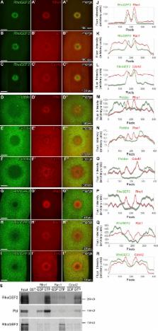
          During wound repair, Rho GTPases form dynamic spatial and temporal patterns surrounding the wound and coordinate the cytoskeletal response. Nakamura et al. show that Rho GTPase arrays form in response to prepatterning by RhoGEFs, which depends on annexin B9.

          Abstract

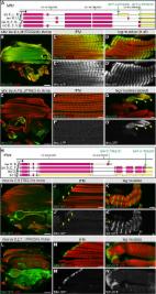
          Like tissues, single cells are subjected to continual stresses and damage. As such, cells have a robust wound repair mechanism comprised of dynamic membrane resealing and cortical cytoskeletal remodeling. One group of proteins, the Rho family of small guanosine triphosphatases (GTPases), is critical for this actin and myosin cytoskeletal response in which they form distinct dynamic spatial and temporal patterns/arrays surrounding the wound. A key mechanistic question, then, is how these GTPase arrays are formed. Here, we show that in the Drosophila melanogaster cell wound repair model Rho GTPase arrays form in response to prepatterning by Rho guanine nucleotide exchange factors (RhoGEFs), a family of proteins involved in the activation of small GTPases. Furthermore, we show that Annexin B9, a member of a class of proteins associated with the membrane resealing, is involved in an early, Rho family–independent, actin stabilization that is integral to the formation of one RhoGEF array. Thus, Annexin proteins may link membrane resealing to cytoskeletal remodeling processes in single cell wound repair.

          Related collections

          Most cited references31

          • Record: found
          • Abstract: found
          • Article: not found

          Multiple Forces Contribute to Cell Sheet Morphogenesis for Dorsal Closure in Drosophila

          The molecular and cellular bases of cell shape change and movement during morphogenesis and wound healing are of intense interest and are only beginning to be understood. Here, we investigate the forces responsible for morphogenesis during dorsal closure with three approaches. First, we use real-time and time-lapsed laser confocal microscopy to follow actin dynamics and document cell shape changes and tissue movements in living, unperturbed embryos. We label cells with a ubiquitously expressed transgene that encodes GFP fused to an autonomously folding actin binding fragment from fly moesin. Second, we use a biomechanical approach to examine the distribution of stiffness/tension during dorsal closure by following the response of the various tissues to cutting by an ultraviolet laser. We tested our previous model (Young, P.E., A.M. Richman, A.S. Ketchum, and D.P. Kiehart. 1993. Genes Dev. 7:29–41) that the leading edge of the lateral epidermis is a contractile purse-string that provides force for dorsal closure. We show that this structure is under tension and behaves as a supracellular purse-string, however, we provide evidence that it alone cannot account for the forces responsible for dorsal closure. In addition, we show that there is isotropic stiffness/tension in the amnioserosa and anisotropic stiffness/tension in the lateral epidermis. Tension in the amnioserosa may contribute force for dorsal closure, but tension in the lateral epidermis opposes it. Third, we examine the role of various tissues in dorsal closure by repeated ablation of cells in the amnioserosa and the leading edge of the lateral epidermis. Our data provide strong evidence that both tissues appear to contribute to normal dorsal closure in living embryos, but surprisingly, neither is absolutely required for dorsal closure. Finally, we establish that the Drosophila epidermis rapidly and reproducibly heals from both mechanical and ultraviolet laser wounds, even those delivered repeatedly. During healing, actin is rapidly recruited to the margins of the wound and a newly formed, supracellular purse-string contracts during wound healing. This result establishes the Drosophila embryo as an excellent system for the investigation of wound healing. Moreover, our observations demonstrate that wound healing in this insect epidermal system parallel wound healing in vertebrate tissues in situ and vertebrate cells in culture (for review see Kiehart, D.P. 1999. Curr. Biol. 9:R602–R605).
            Bookmark
            • Record: found
            • Abstract: found
            • Article: found
            Is Open Access

            A genome-wide resource for the analysis of protein localisation in Drosophila

            The Drosophila genome contains >13000 protein-coding genes, the majority of which remain poorly investigated. Important reasons include the lack of antibodies or reporter constructs to visualise these proteins. Here, we present a genome-wide fosmid library of 10000 GFP-tagged clones, comprising tagged genes and most of their regulatory information. For 880 tagged proteins, we created transgenic lines, and for a total of 207 lines, we assessed protein expression and localisation in ovaries, embryos, pupae or adults by stainings and live imaging approaches. Importantly, we visualised many proteins at endogenous expression levels and found a large fraction of them localising to subcellular compartments. By applying genetic complementation tests, we estimate that about two-thirds of the tagged proteins are functional. Moreover, these tagged proteins enable interaction proteomics from developing pupae and adult flies. Taken together, this resource will boost systematic analysis of protein expression and localisation in various cellular and developmental contexts. DOI: http://dx.doi.org/10.7554/eLife.12068.001
              Bookmark
              • Record: found
              • Abstract: found
              • Article: not found

              Concentric zones of active RhoA and Cdc42 around single cell wounds

              Rho GTPases control many cytoskeleton-dependent processes, but how they regulate spatially distinct features of cytoskeletal function within a single cell is poorly understood. Here, we studied active RhoA and Cdc42 in wounded Xenopus oocytes, which assemble and close a dynamic ring of actin filaments (F-actin) and myosin-2 around wound sites. RhoA and Cdc42 are rapidly activated around wound sites in a calcium-dependent manner and segregate into distinct, concentric zones around the wound, with active Cdc42 in the approximate middle of the F-actin array and active RhoA on the interior of the array. These zones form before F-actin accumulation, and then move in concert with the closing array. Microtubules and F-actin are required for normal zone organization and dynamics, as is crosstalk between RhoA and Cdc42. Each of the zones makes distinct contributions to the organization and function of the actomyosin wound array. We propose that similar rho activity zones control related processes such as cytokinesis.
                Bookmark

                Author and article information

                Journal
                J Cell Biol
                J. Cell Biol
                jcb
                jcb
                The Journal of Cell Biology
                The Rockefeller University Press
                0021-9525
                1540-8140
                4 December 2017
                : 216
                : 12
                : 3959-3969
                Affiliations
                [1]Basic Sciences Division, Fred Hutchinson Cancer Research Center, Seattle, WA
                Author notes
                Correspondence to Susan M. Parkhurst: susanp@ 123456fredhutch.org
                Author information
                http://orcid.org/0000-0001-7879-3176
                http://orcid.org/0000-0001-5806-9930
                Article
                201704145
                10.1083/jcb.201704145
                5716286
                28923977
                0006f2cd-dd1b-43ea-977b-5cc1561284e1
                © 2017 Nakamura et al.

                This article is distributed under the terms of an Attribution–Noncommercial–Share Alike–No Mirror Sites license for the first six months after the publication date (see http://www.rupress.org/terms/). After six months it is available under a Creative Commons License (Attribution–Noncommercial–Share Alike 4.0 International license, as described at https://creativecommons.org/licenses/by-nc-sa/4.0/).

                History
                : 24 April 2017
                : 17 August 2017
                : 24 August 2017
                Funding
                Funded by: National Institutes of Health, DOI http://dx.doi.org/10.13039/100000002;
                Award ID: GM111635
                Categories
                Research Articles
                Report
                38
                40
                10

                Cell biology
                Cell biology

                Comments

                Comment on this article