75
views
0
recommends
+1 Recommend
0 collections
    0
    shares
      • Record: found
      • Abstract: found
      • Article: not found

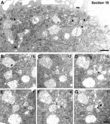
      In macrophages, HIV-1 assembles into an intracellular plasma membrane domain containing the tetraspanins CD81, CD9, and CD53

      research-article

      Read this article at

      ScienceOpenPublisherPMC
      Bookmark
          There is no author summary for this article yet. Authors can add summaries to their articles on ScienceOpen to make them more accessible to a non-specialist audience.

          Abstract

          In macrophages, HIV-1 has been shown to bud into intracellular structures that contain the late endosome marker CD63. We show that these organelles are not endosomes, but an internally sequestered plasma membrane domain. Using immunofluorescence microscopy and immunoelectron microscopy, we find that HIV-1 buds into a compartment that contains the tetraspanins CD81, CD9, and CD53. On uninfected macrophages, these proteins are seen at the cell surface and in intracellular vacuole-like structures with a complex content of vesicles and interconnected membranes that lack endosome markers, including CD63. Significantly, these structures are accessible to small tracers (horseradish peroxidase or ruthenium red) applied to cells at 4°C, indicating that they are connected to the cell surface. HIV assembles on, and accumulates within, these intracellular compartments. Furthermore, CD63 is recruited to the virus-containing structures and incorporated into virions. These results indicate that, in macrophages, HIV-1 exploits a previously undescribed intracellular plasma membrane domain to assemble infectious particles.

          Related collections

          Most cited references40

          • Record: found
          • Abstract: found
          • Article: not found

          Selective enrichment of tetraspan proteins on the internal vesicles of multivesicular endosomes and on exosomes secreted by human B-lymphocytes.

          Association of major histocompatibility complex (MHC) class II molecules with peptides occurs in a series of endocytic vacuoles, termed MHC class II-enriched compartments (MIICs). Morphological criteria have defined several types of MIICs, including multivesicular MIICs, which are composed of 50-60-nm vesicles surrounded by a limiting membrane. Multivesicular MIICs can fuse with the plasma membrane, thereby releasing their internal vesicles into the extracellular space. The externalized vesicles, termed exosomes, carry MHC class II and can stimulate T-cells in vitro. In this study, we show that exosomes are enriched in the co-stimulatory molecule CD86 and in several tetraspan proteins, including CD37, CD53, CD63, CD81, and CD82. Interestingly, subcellular localization of these molecules revealed that they were concentrated on the internal membranes of multivesicular MIICs. In contrast to the tetraspans, other membrane proteins of MIICs, such as HLA-DM, Lamp-1, and Lamp-2, were mainly localized to the limiting membrane and were hardly detectable on the internal membranes of MIICs nor on exosomes. Because internal vesicles of multivesicular MIICs are thought to originate from inward budding of the limiting membrane, the differential distribution of membrane proteins on the internal and limiting membranes of MIICs has to be driven by active protein sorting.
            Bookmark
            • Record: found
            • Abstract: found
            • Article: not found

            Recruitment of HIV and its receptors to dendritic cell-T cell junctions.

            Monocyte-derived dendritic cells (MDDCs) can efficiently bind and transfer HIV infectivity without themselves becoming infected. Using live-cell microscopy, we found that HIV was recruited to sites of cell contact in MDDCs. Analysis of conjugates between MDDCs and T cells revealed that, in the absence of antigen-specific signaling, the HIV receptors CD4, CCR5, and CXCR4 on the T cell were recruited to the interface while the MDDCs concentrated HIV to the same region. We propose that contact between dendritic cells and T cells facilitates transmission of HIV by locally concentrating virus, receptor, and coreceptor during the formation of an infectious synapse.
              Bookmark
              • Record: found
              • Abstract: found
              • Article: not found

              Structural basis for targeting HIV-1 Gag proteins to the plasma membrane for virus assembly.

              During the late phase of HIV type 1 (HIV-1) replication, newly synthesized retroviral Gag proteins are targeted to the plasma membrane of most hematopoietic cell types, where they colocalize at lipid rafts and assemble into immature virions. Membrane binding is mediated by the matrix (MA) domain of Gag, a 132-residue polypeptide containing an N-terminal myristyl group that can adopt sequestered and exposed conformations. Although exposure is known to promote membrane binding, the mechanism by which Gag is targeted to specific membranes has yet to be established. Recent studies have shown that phosphatidylinositol (PI) 4,5-bisphosphate [PI(4,5)P(2)], a factor that regulates localization of cellular proteins to the plasma membrane, also regulates Gag localization and assembly. Here we show that PI(4,5)P(2) binds directly to HIV-1 MA, inducing a conformational change that triggers myristate exposure. Related phosphatidylinositides PI, PI(3)P, PI(4)P, PI(5)P, and PI(3,5)P(2) do not bind MA with significant affinity or trigger myristate exposure. Structural studies reveal that PI(4,5)P(2) adopts an "extended lipid" conformation, in which the inositol head group and 2'-fatty acid chain bind to a hydrophobic cleft, and the 1'-fatty acid and exposed myristyl group bracket a conserved basic surface patch previously implicated in membrane binding. Our findings indicate that PI(4,5)P(2) acts as both a trigger of the myristyl switch and a membrane anchor and suggest a potential mechanism for targeting Gag to membrane rafts.
                Bookmark

                Author and article information

                Journal
                J Cell Biol
                jcb
                The Journal of Cell Biology
                The Rockefeller University Press
                0021-9525
                1540-8140
                23 April 2007
                : 177
                : 2
                : 329-341
                Affiliations
                [1 ]Cell Biology Unit, Medical Research Council Laboratory for Molecular Cell Biology, and [2 ]Department of Biochemistry and Molecular Biology, University College London, London WC1E 6BT, England, UK
                Author notes

                Correspondence to Mark Marsh: m.marsh@ 123456ucl.ac.uk

                Article
                200609050
                10.1083/jcb.200609050
                2064140
                17438075
                05a16faa-c775-4819-ba61-9cb11f52eb98
                Copyright © 2007, The Rockefeller University Press
                History
                : 8 September 2006
                : 20 March 2007
                Categories
                Research Articles
                Article

                Cell biology
                Cell biology

                Comments

                Comment on this article