28
views
0
recommends
+1 Recommend
0 collections
    0
    shares
      • Record: found
      • Abstract: found
      • Article: found
      Is Open Access

      Effects of conventional immunosuppressive treatment on CD244+ (CD28null) and FOXP3+ T cells in the inflamed muscle of patients with polymyositis and dermatomyositis

      research-article

      Read this article at

      Bookmark
          There is no author summary for this article yet. Authors can add summaries to their articles on ScienceOpen to make them more accessible to a non-specialist audience.

          Abstract

          Background

          T-cell infiltrates may persist in muscle tissue of polymyositis (PM) and dermatomyositis (DM) patients despite aggressive immunosuppressive treatment. Here, we investigated to what extent persistent T cells in affected muscle were FOXP3+, a marker for regulatory T cells (Tregs), or CD244+, a marker for CD28null T cells, and whether their presence correlated to clinical outcome. The sensitivity of CD28null T cells towards glucocorticoid and Treg-mediated immunosuppression was also investigated.

          Methods

          Muscle biopsies from 16 newly diagnosed or untreated patients with PM/DM were investigated by immunohistochemistry for expression of CD3, FOXP3 and CD244 before and after treatment with glucocorticoids and immunosuppressive agents. For clinical evaluation, serum levels of creatine kinase, muscle performance (FI and MMT8), disease activity (MITAX) and disability (HAQ) were measured. In vitro suppressive effects of glucocorticoids and Tregs on T-cell activation were measured by CD69 upregulation.

          Results

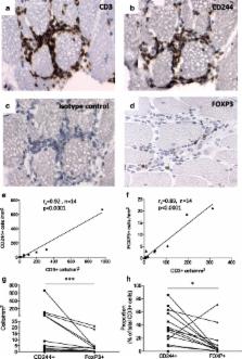
          Before treatment, CD244+ cells were present at higher proportions compared to FOXP3+ cells in the inflamed muscle. Following treatment, FOXP3+ cell numbers decreased while CD244+ cells persisted. Patients with impaired muscle function (<75 % FI) post-treatment had higher levels of CD244+ cells in the follow-up biopsy compared to those with FI >75 %. MITAX and HAQ correlated with the number of CD244+ cells post-treatment. CD4+CD28null T cells displayed lower sensitivity towards both glucocorticoid and Treg-mediated immunosuppression in vitro compared to their CD28+ counterparts.

          Conclusions

          Poor outcome in patients with myositis following immunosuppressive therapy was linked to persistence of CD244+ (CD28null) T cells in muscle tissue, suggesting their resistance against immunosuppression. A relative loss of regulatory T cells could also contribute to poor clinical outcome given their recently ascribed role in muscle tissue regeneration.

          Related collections

          Most cited references40

          • Record: found
          • Abstract: found
          • Article: not found

          Regulatory interactions between muscle and the immune system during muscle regeneration.

          Recent discoveries reveal complex interactions between skeletal muscle and the immune system that regulate muscle regeneration. In this review, we evaluate evidence that indicates that the response of myeloid cells to muscle injury promotes muscle regeneration and growth. Acute perturbations of muscle activate a sequence of interactions between muscle and inflammatory cells. The initial inflammatory response is a characteristic Th1 inflammatory response, first dominated by neutrophils and subsequently by CD68(+) M1 macrophages. M1 macrophages can propagate the Th1 response by releasing proinflammatory cytokines and cause further tissue damage through the release of nitric oxide. Myeloid cells in the early Th1 response stimulate the proliferative phase of myogenesis through mechanisms mediated by TNF-alpha and IL-6; experimental prolongation of their presence is associated with delayed transition to the early differentiation stage of myogenesis. Subsequent invasion by CD163(+)/CD206(+) M2 macrophages attenuates M1 populations through the release of anti-inflammatory cytokines, including IL-10. M2 macrophages play a major role in promoting growth and regeneration; their absence greatly slows muscle growth following injury or modified use and inhibits muscle differentiation and regeneration. Chronic muscle injury leads to profiles of macrophage invasion and function that differ from acute injuries. For example, mdx muscular dystrophy yields invasion of muscle by M1 macrophages, but their early invasion is accompanied by a subpopulation of M2a macrophages. M2a macrophages are IL-4 receptor(+)/CD206(+) cells that reduce cytotoxicity of M1 macrophages. Subsequent invasion of dystrophic muscle by M2c macrophages is associated with progression of the regenerative phase in pathophysiology. Together, these findings show that transitions in macrophage phenotype are an essential component of muscle regeneration in vivo following acute or chronic muscle damage.
            Bookmark
            • Record: found
            • Abstract: found
            • Article: not found

            Glucocorticoids in T cell development and function*.

            Glucocorticoids are small lipophilic compounds that mediate their many biological effects by binding an intracellular receptor (GR) that, in turn, translocates to the nucleus and directly or indirectly regulates gene transcription. Perhaps the most recognized biologic effect of glucocorticoids on peripheral T cells is immunosuppression, which is due to inhibition of expression of a wide variety of activationinduced gene products. Glucocorticoids have also been implicated in Th lineage development (favoring the generation of Th2 cells) and, by virtue of their downregulation of fasL expression, the inhibition of activation-induced T cell apoptosis. Glucocorticoids are also potent inducers of apoptosis, and even glucocorticoid concentrations achieved during a stress response can cause the death of CD4(+)CD8(+ )thymocytes. Perhaps surprisingly, thymic epithelial cells produce glucocorticoids, and based upon in vitro and in vivo studies of T cell development it has been proposed that these locally produced glucocorticoids participate in antigen-specific thymocyte development by inhibiting activation-induced gene transcription and thus increasing the TCR signaling thresholds required to promote positive and negative selection. It is anticipated that studies in animals with tissue-specific GR-deficiency will further elucide how glucocorticoids affect T cell development and function.
              Bookmark
              • Record: found
              • Abstract: found
              • Article: not found

              Monoclonal T-cell proliferation and plaque instability in acute coronary syndromes.

              Unstable angina (UA) is associated with systemic inflammation and with expansion of interferon-gamma-producing T lymphocytes. The cause of T-cell activation and the precise role of activated T cells in plaque instability are not understood. Peripheral blood T cells from 34 patients with stable angina and 34 patients with UA were compared for the distribution of functional T-cell subsets by flow cytometric analysis. Clonality within the T-cell compartment was identified by T-cell receptor spectrotyping and subsequent sequencing. Tissue-infiltrating T cells were examined in extracts from coronary arteries containing stable or unstable plaque. The subset of CD4(+)CD28(null) T cells was expanded in patients with UA and infrequent in patients with stable angina (median frequencies: 10.8% versus 1.5%, P<0.001). CD4(+)CD28(null) T cells included a large monoclonal population, with 59 clonotypes isolated from 20 UA patients. T-cell clonotypes from different UA patients used antigen receptors with similar sequences. T-cell receptor sequences derived from monoclonal T-cell populations were detected in the culprit but not in the nonculprit lesion of a patient with fatal myocardial infarction. UA is associated with the emergence of monoclonal T-cell populations, analogous to monoclonal gammopathy of unknown significance. Shared T-cell receptor sequences in clonotypes of different patients implicate chronic stimulation by a common antigen, for example, persistent infection. The unstable plaque but not the stable plaque is invaded by clonally expanded T cells, suggesting a direct involvement of these lymphocytes in plaque disruption.
                Bookmark

                Author and article information

                Contributors
                jayesh.pandya05@gmail.com
                Ingela.Loell@ki.se
                ms_hossain77@yahoo.com
                mei.zong@ki.se
                helene.Alexanderson@karolinska.se
                sukanya.raghavan@microbio.gu.se
                Ingrid.Lundberg@ki.se
                +46 (0) 8 517 75609 , vivianne.malmstrom@ki.se
                Journal
                Arthritis Res Ther
                Arthritis Res. Ther
                Arthritis Research & Therapy
                BioMed Central (London )
                1478-6354
                1478-6362
                1 April 2016
                1 April 2016
                2016
                : 18
                : 80
                Affiliations
                [ ]Rheumatology Unit, Department of Medicine, Center for Molecular Medicine, Karolinska University Hospital, Solna, Karolinska Institutet, Stockholm, Sweden
                [ ]Department of NVS, Division of Physiotherapy, Solna, Karolinska Institutet, Huddinge and Physiotherapy Clinic, Karolinska University Hospital, Stockholm, Sweden
                [ ]Present address: Department of Microbiology and Immunology, Institute for Biomedicine, Gothenburg University, Gothenburg, Sweden
                Article
                974
                10.1186/s13075-016-0974-5
                4818535
                27039301
                076fe8df-b64c-4866-bd93-4090f326d519
                © Pandya et al. 2016

                Open AccessThis article is distributed under the terms of the Creative Commons Attribution 4.0 International License ( http://creativecommons.org/licenses/by/4.0/), which permits unrestricted use, distribution, and reproduction in any medium, provided you give appropriate credit to the original author(s) and the source, provide a link to the Creative Commons license, and indicate if changes were made. The Creative Commons Public Domain Dedication waiver ( http://creativecommons.org/publicdomain/zero/1.0/) applies to the data made available in this article, unless otherwise stated.

                History
                : 21 December 2015
                : 14 March 2016
                Funding
                Funded by: AutoCure
                Award ID: LSH-018661
                Award Recipient :
                Funded by: Swedish Research Council
                Funded by: The Swedish Rheumatism Association
                Funded by: King Gustaf V 80 Year Foundation
                Funded by: Karolinska Institutet foundation (KID) and through the regional agreement on medical training and clinical research (ALF) between Stockholm County Council and Karolinska Institutet.
                Funded by: FundRef http://dx.doi.org/http://dx.doi.org/10.13039/501100004359, Vetenskapsrådet;
                Funded by: King Gustaf V:s 80-year Foundation
                Categories
                Research Article
                Custom metadata
                © The Author(s) 2016

                Orthopedics
                t-lymphocyte,myositis,treg cells,glucocorticoids,inflammation
                Orthopedics
                t-lymphocyte, myositis, treg cells, glucocorticoids, inflammation

                Comments

                Comment on this article