83
views
0
recommends
+1 Recommend
0 collections
    0
    shares
      • Record: found
      • Abstract: found
      • Article: found
      Is Open Access

      Linking ER Stress to Autophagy: Potential Implications for Cancer Therapy

      review-article

      Read this article at
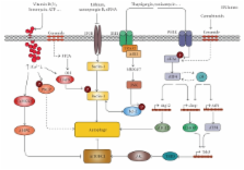
      Bookmark
          There is no author summary for this article yet. Authors can add summaries to their articles on ScienceOpen to make them more accessible to a non-specialist audience.

          Abstract

          Different physiological and pathological conditions can perturb protein folding in the endoplasmic reticulum, leading to a condition known as ER stress. ER stress activates a complex intracellular signal transduction pathway, called unfolded protein response (UPR). The UPR is tailored essentially to reestablish ER homeostasis also through adaptive mechanisms involving the stimulation of autophagy. However, when persistent, ER stress can switch the cytoprotective functions of UPR and autophagy into cell death promoting mechanisms. Recently, a variety of anticancer therapies have been linked to the induction of ER stress in cancer cells, suggesting that strategies devised to stimulate its prodeath function or block its prosurvival function, could be envisaged to improve their tumoricidial action. A better understanding of the molecular mechanisms that determine the final outcome of UPR and autophagy activation by chemotherapeutic agents, will offer new opportunities to improve existing cancer therapies as well as unravel novel targets for cancer treatment.

          Related collections

          Most cited references148

          • Record: found
          • Abstract: found
          • Article: not found

          Autophagy is activated for cell survival after endoplasmic reticulum stress.

          Eukaryotic cells deal with accumulation of unfolded proteins in the endoplasmic reticulum (ER) by the unfolded protein response, involving the induction of molecular chaperones, translational attenuation, and ER-associated degradation, to prevent cell death. Here, we found that the autophagy system is activated as a novel signaling pathway in response to ER stress. Treatment of SK-N-SH neuroblastoma cells with ER stressors markedly induced the formation of autophagosomes, which were recognized at the ultrastructural level. The formation of green fluorescent protein (GFP)-LC3-labeled structures (GFP-LC3 "dots"), representing autophagosomes, was extensively induced in cells exposed to ER stress with conversion from LC3-I to LC3-II. In IRE1-deficient cells or cells treated with c-Jun N-terminal kinase (JNK) inhibitor, the autophagy induced by ER stress was inhibited, indicating that the IRE1-JNK pathway is required for autophagy activation after ER stress. In contrast, PERK-deficient cells and ATF6 knockdown cells showed that autophagy was induced after ER stress in a manner similar to the wild-type cells. Disturbance of autophagy rendered cells vulnerable to ER stress, suggesting that autophagy plays important roles in cell survival after ER stress.
            Bookmark
            • Record: found
            • Abstract: found
            • Article: not found

            The role of autophagy in cancer development and response to therapy.

            Autophagy is a process in which subcellular membranes undergo dynamic morphological changes that lead to the degradation of cellular proteins and cytoplasmic organelles. This process is an important cellular response to stress or starvation. Many studies have shed light on the importance of autophagy in cancer, but it is still unclear whether autophagy suppresses tumorigenesis or provides cancer cells with a rescue mechanism under unfavourable conditions. What is the present state of our knowledge about the role of autophagy in cancer development, and in response to therapy? And how can the autophagic process be manipulated to improve anticancer therapeutics?
              Bookmark
              • Record: found
              • Abstract: found
              • Article: not found

              CHOP is implicated in programmed cell death in response to impaired function of the endoplasmic reticulum.

              Cellular stress, particularly in response to toxic and metabolic insults that perturb function of the endoplasmic reticulum (ER stress), is a powerful inducer of the transcription factor CHOP. The role of CHOP in the response of cells to injury associated with ER stress was examined in a murine deficiency model obtained by homologous recombination at the chop gene. Compared with the wild type, mouse embryonic fibroblasts (MEFs) derived from chop -/- animals exhibited significantly less programmed cell death when challenged with agents that perturb ER function. A similar deficit in programmed cells death in response to ER stress was also observed in MEFs that lack CHOP's major dimerization partner, C/EBPbeta, implicating the CHOP-C/EBP pathway in programmed cell death. An animal model for studying the effects of chop on the response to ER stress was developed. It entailed exposing mice with defined chop genotypes to a single sublethal intraperitoneal injection of tunicamycin and resulted in a severe illness characterized by transient renal insufficiency. In chop +/+ and chop +/- mice this was associated with the early expression of CHOP in the proximal tubules followed by the development of a histological picture similar to the human condition known as acute tubular necrosis, a process that resolved by cellular regeneration. In the chop -/- animals, in spite of the severe impairment in renal function, evidence of cellular death in the kidney was reduced compared with the wild type. The proximal tubule epithelium of chop -/- animals exhibited fourfold lower levels of TUNEL-positive cells (a marker for programmed cell death), and significantly less evidence for subsequent regeneration. CHOP therefore has a role in the induction of cell death under conditions associated with malfunction of the ER and may also have a role in cellular regeneration under such circumstances.
                Bookmark

                Author and article information

                Journal
                Int J Cell Biol
                IJCB
                International Journal of Cell Biology
                Hindawi Publishing Corporation
                1687-8876
                1687-8884
                2010
                17 January 2010
                : 2010
                : 930509
                Affiliations
                1Department of Molecular Cell Biology, Cell Death Research and Therapy Laboratory, Faculty of Medicine, Catholic University of Leuven, 3000 Leuven, Belgium
                2Department of Biochemistry and Molecular Biology I, School of Biology, Complutense University, 28040 Madrid, Spain
                3Centro de Investigación Biomédica en Red sobre Enfermedades Neurodegenerativas (CIBERNED), 28040 Madrid, Spain
                Author notes
                *Guillermo Velasco: gvd@ 123456bbm1.ucm.es and

                Academic Editor: Afshin Samali

                Article
                10.1155/2010/930509
                2817393
                20145727
                0ef094e0-1a13-4cc5-b06b-bc06520259a1
                Copyright © 2010 Tom Verfaillie et al.

                This is an open access article distributed under the Creative Commons Attribution License, which permits unrestricted use, distribution, and reproduction in any medium, provided the original work is properly cited.

                History
                : 23 July 2009
                : 25 September 2009
                : 19 October 2009
                Categories
                Review Article

                Cell biology
                Cell biology

                Comments

                Comment on this article