9
views
0
recommends
+1 Recommend
0 collections
    0
    shares
      • Record: found
      • Abstract: found
      • Article: found
      Is Open Access

      Multiple Ligament Reconstructions of the Knee and Posterolateral Corner

      brief-report
      , M.S. a , b , , M.D. a , , P.T., O.C.S. c , , M.D., Ph.D. a ,
      Arthroscopy Techniques
      Elsevier

      Read this article at

      Bookmark
          There is no author summary for this article yet. Authors can add summaries to their articles on ScienceOpen to make them more accessible to a non-specialist audience.

          Abstract

          Injuries to the knee involving multiple ligaments occur in a variety of circumstances and require careful assessment and planning. A wide constellation of injuries can occur with causes sufficiently traumatic to produce bicruciate ligament deficiency, and this technical report will describe diagnosis, treatment and rehabilitation for a knee dislocation with lateral injury (KD-III-L on the Schenk classification). Reconstruction in the acute setting is preferred, with anatomic-based, single-bundle anterior cruciate ligament reconstruction, double-bundle posterior cruciate ligament reconstruction, and anatomic reconstruction of the posterolateral corner using two grafts for the 3 primary posterolateral corner stabilizers. Tunnel orientation to prevent convergence and sequence of graft tensioning and fixation are discussed as well. Successful outcomes have been achieved using these anatomic-based reconstruction techniques along with appropriate rehabilitation and bracing.

          Technique Video

          Video 1

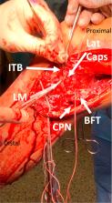
          Repair of bicruciate ligament injury with grade III posterolateral corner tear. Examination under anesthesia is consistent with KD-III-L injury. Dissection of biceps femoris tendon (BFT) and common peroneal nerve (CPN) is performed through a lateral hockey stick incision. A fibular tunnel is drilled first, then a tibial tunnel for the popliteus tendon (PLT) and popliteofibular ligament (PLT). Femoral tunnels for the PLT and fibular collateral ligament (FCL) are drilled, oriented 35°anteromedially. Bone-patellar tendon-bone autograft is then harvested. After diagnostic arthroscopy, the PCL anterolateral bundle (ALB) and posteromedial bundle (PMB) tunnels are drilled, then the anterior cruciate ligament (ACL) femoral tunnel. A tibial guide pin is placed, confirmed with fluoroscopy, then a tunnel reamed and a large smoother passed. The ACL tibial tunnel is then reamed. The PMB graft of the PCL is passed first and secured, then the ALB. The ends of both bundles are passed down the tibial tunnel using the smoother. The ACL femoral graft is pulled into place and secured. The femoral ends of the FCL and PLT grafts are then secured. The FCL graft is passed under the iliotibial band (ITB) and through the fibular head tunnel. PCL grafts are then fixed on the anterior tibial cortex with screws and washers; ALB in 90° flexion and PMB in extension. This eliminated the posterior drawer. The ACL graft is then secured on the anterior tibia. The FCL graft is then secured in the fibular head tunnel with a screw at the anterior aperture. The PLT and PFL grafts are then passed from posterior to anterior through the tibial PLC tunnel and secured with a screw. Suture anchors are used to repair the lateral capsule and biceps femoris tendon, and the ITB repaired. Closure completes the procedure.

          Related collections

          Most cited references37

          • Record: found
          • Abstract: found
          • Article: found

          Blood flow restriction training in clinical musculoskeletal rehabilitation: a systematic review and meta-analysis.

          Low-load exercise training with blood flow restriction (BFR) can increase muscle strength and may offer an effective clinical musculoskeletal (MSK) rehabilitation tool. The aim of this review was to systematically analyse the evidence regarding the effectiveness of this novel training modality in clinical MSK rehabilitation.
            Bookmark
            • Record: found
            • Abstract: found
            • Article: not found

            Quadriceps activation following knee injuries: a systematic review.

            Arthrogenic muscle inhibition is an important underlying factor in persistent quadriceps muscle weakness after knee injury or surgery. To determine the magnitude and prevalence of volitional quadriceps activation deficits after knee injury. Web of Science database. Eligible studies involved human participants and measured quadriceps activation using either twitch interpolation or burst superimposition on patients with knee injuries or surgeries such as anterior cruciate ligament deficiency (ACLd), anterior cruciate ligament reconstruction (ACLr), and anterior knee pain (AKP). Means, measures of variability, and prevalence of quadriceps activation (QA) failure (<95%) were recorded for experiments involving ACLd (10), ACLr (5), and AKP (3). A total of 21 data sets from 18 studies were initially identified. Data from 3 studies (1 paper reporting data for both ACLd and ACLr, 1 on AKP, and the postarthroscopy paper) were excluded from the primary analyses because only graphical data were reported. Of the remaining 17 data sets (from 15 studies), weighted mean QA in 352 ACLd patients was 87.3% on the involved side, 89.1% on the uninvolved side, and 91% in control participants. The QA failure prevalence ranged from 0% to 100%. Weighted mean QA in 99 total ACLr patients was 89.2% on the involved side, 84% on the uninvolved side, and 98.5% for the control group, with prevalence ranging from 0% to 71%. Thirty-eight patients with AKP averaged 78.6% on the involved side and 77.7% on the contralateral side. Bilateral QA failure was commonly reported in patients. Quadriceps activation failure is common in patients with ACLd, ACLr, and AKP and is often observed bilaterally.
              Bookmark
              • Record: found
              • Abstract: found
              • Article: not found

              Increased risk of revision with hamstring tendon grafts compared with patellar tendon grafts after anterior cruciate ligament reconstruction: a study of 12,643 patients from the Norwegian Cruciate Ligament Registry, 2004-2012.

              The graft choice for anterior cruciate ligament reconstruction (ACLR) is controversial. Hamstring tendon (HT) autografts and patellar tendon (PT) autografts are the most common grafts used and have shown similar subjective and objective outcomes.
                Bookmark

                Author and article information

                Contributors
                Journal
                Arthrosc Tech
                Arthrosc Tech
                Arthroscopy Techniques
                Elsevier
                2212-6287
                12 April 2021
                May 2021
                12 April 2021
                : 10
                : 5
                : e1269-e1280
                Affiliations
                [a ]Twin Cities Orthopedics, Edina-Crosstown, Edina, Minnesota
                [b ]Georgetown University School of Medicine, Washington, District of Columbia
                [c ]Training Haus, Twin Cities Orthopedics, Eagan–Viking Lakes, Minnesota
                Author notes
                []Address correspondence to Robert F. LaPrade, M.D., Ph.D., Twin Cities Orthopedics, Edina-Crosstown, 4010 W 65th St., Edina, MN 55435-1706, U.S.A. laprademdphd@ 123456gmail.com
                Article
                S2212-6287(21)00030-X
                10.1016/j.eats.2021.01.024
                8185621
                34141542
                12098e48-a2f5-429a-8872-33a2ad7a917d
                © 2021 by the Arthroscopy Association of North America. Published by Elsevier.

                This is an open access article under the CC BY-NC-ND license (http://creativecommons.org/licenses/by-nc-nd/4.0/).

                History
                : 16 December 2020
                : 29 January 2021
                Categories
                Technical Note

                Comments

                Comment on this article