69
views
0
recommends
+1 Recommend
0 collections
    0
    shares
      • Record: found
      • Abstract: found
      • Article: found
      Is Open Access

      VEGF-A modulates expression of inhibitory checkpoints on CD8 + T cells in tumors

      research-article

      Read this article at

      Bookmark
          There is no author summary for this article yet. Authors can add summaries to their articles on ScienceOpen to make them more accessible to a non-specialist audience.

          Abstract

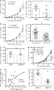
          VEGF-A production in the tumor microenvironment enhances expression of PD-1 and other inhibitory checkpoints involved with CD8 + T cell exhaustion, which can be reversed with anti-VEGF/VEGFR treatment.

          Abstract

          Immune escape is a prerequisite for tumor development. To avoid the immune system, tumors develop different mechanisms, including T cell exhaustion, which is characterized by expression of immune inhibitory receptors, such as PD-1, CTLA-4, Tim-3, and a progressive loss of function. The recent development of therapies targeting PD-1 and CTLA-4 have raised great interest since they induced long-lasting objective responses in patients suffering from advanced metastatic tumors. However, the regulation of PD-1 expression, and thereby of exhaustion, is unclear. VEGF-A, a proangiogenic molecule produced by the tumors, plays a key role in the development of an immunosuppressive microenvironment. We report in the present work that VEGF-A produced in the tumor microenvironment enhances expression of PD-1 and other inhibitory checkpoints involved in CD8 + T cell exhaustion, which could be reverted by anti-angiogenic agents targeting VEGF-A–VEGFR. In view of these results, association of anti-angiogenic molecules with immunomodulators of inhibitory checkpoints may be of particular interest in VEGF-A-producing tumors.

          Related collections

          Most cited references14

          • Record: found
          • Abstract: found
          • Article: not found

          The novel role of tyrosine kinase inhibitor in the reversal of immune suppression and modulation of tumor microenvironment for immune-based cancer therapies.

          In tumor-bearing hosts, myeloid-derived suppressor cells (MDSC) and T regulatory cells (Treg) play important roles in immune suppression, the reversal of which is vitally important for the success of immune therapy. We have shown that ckit ligand is required for MDSC accumulation and Treg development. We hypothesized that sunitinib malate, a receptor tyrosine kinase inhibitor, could reverse MDSC-mediated immune suppression and modulate the tumor microenvironment, thereby improving the efficacy of immune-based therapies. Treatment with sunitinib decreased the number of MDSC and Treg in advanced tumor-bearing animals. Furthermore, it not only reduced the suppressive function of MDSCs but also prevented tumor-specific T-cell anergy and Treg development. Interestingly, sunitinib treatment resulted in reduced expression of interleukin (IL)-10, transforming growth factor-beta, and Foxp3 but enhanced expression of Th1 cytokine IFN-gamma and increased CTL responses in isolated tumor-infiltrating leukocytes. A significantly higher percentage and infiltration of CD8 and CD4 cells was detected in tumors of sunitinib-treated mice when compared with control-treated mice. More importantly, the expression of negative costimulatory molecules CTLA4 and PD-1 in both CD4 and CD8 T cells, and PDL-1 expression on MDSC and plasmacytoid dendritic cells, was also significantly decreased by sunitinib treatment. Finally, sunitinib in combination with our immune therapy protocol (IL-12 and 4-1BB activation) significantly improves the long-term survival rate of large tumor-bearing mice. These data suggest that sunitinib can be used to reverse immune suppression and as a potentially useful adjunct for enhancing the efficacy of immune-based cancer therapy for advanced malignancies.
            Bookmark
            • Record: found
            • Abstract: found
            • Article: not found

            Sunitinib reverses type-1 immune suppression and decreases T-regulatory cells in renal cell carcinoma patients.

            Immune dysfunction is well documented in renal cell carcinoma (RCC) patients and likely contributes to tumor evasion. This dysfunction includes a shift from a type-1 to a type-2 T-cell cytokine response and enhanced T-regulatory (Treg) cell expression. Given the antitumor activity of select tyrosine kinase inhibitors such as sunitinib in metastatic RCC (mRCC) patients, it is relevant to assess their effect on the immune system. Type-1 (IFNgamma) and type-2 (interleukin-4) responses were assessed in T cells at baseline and day 28 of treatment with sunitinib (50 mg/d) by measuring intracellular cytokines after in vitro stimulation with anti-CD3/anti-CD28 antibodies. After one cycle of treatment, there was a significant increase in the percentage of IFNgamma-producing T cells (CD3(+), P < 0.001; CD3(+)CD4(+), P = 0.001), a reduction in interleukin-4 production (CD3(+) cells, P = 0.05), and a diminished type-2 bias (P = 0.005). The increase in type-1 response may be partly related to modulation of Treg cells. The increased percentage of Treg cells noted in mRCC patients over healthy donors (P = 0.001) was reduced after treatment, although not reaching statistical significance. There was, however, an inverse correlation between the increase in type-1 response after two cycles of treatment and a decrease in the percentage of Treg cells (r = -0.64, P = 0.01). In vitro studies suggest that the effects of sunitinib on Treg cells are indirect. The demonstration that sunitinib improved type-1 T-cell cytokine response in mRCC patients while reducing Treg function provides a basis for the rational combination of sunitinib and immunotherapy in mRCC.
              Bookmark
              • Record: found
              • Abstract: found
              • Article: not found

              Production of vascular endothelial growth factor by human tumors inhibits the functional maturation of dendritic cells.

              Inadequate presentation of tumor antigens by host professional antigen-presenting cells (APCs), including dendritic cells (DCs), is one potential mechanism for the escape of tumors from the host immune system. Here, we show that human cancer cell lines release a soluble factor or factors that dramatically affect DC maturation from precursors without affecting the function of relatively mature DCs. One factor responsible for these effects was identified as vascular endothelial growth factor (VEGF). Thus, VEGF may play a broader role in the pathogenesis of cancer than was previously thought, and therapeutic blockade of VEGF action may improve prospects for immunotherapy as well as inhibit tumor neovasculature.
                Bookmark

                Author and article information

                Journal
                J Exp Med
                J. Exp. Med
                jem
                jem
                The Journal of Experimental Medicine
                The Rockefeller University Press
                0022-1007
                1540-9538
                9 February 2015
                : 212
                : 2
                : 139-148
                Affiliations
                [1 ]INSERM U970, Paris Cardiovascular Research Center, Université Paris-Descartes, Sorbonne Paris Cité, 75015 Paris, France
                [2 ]Service d’immunologie biologique , [3 ]Service d’oncologie médicale , [4 ]Service de chirurgie digestive , [5 ]Service d’hépatogastroentérologie et d’oncologie digestive, Hôpital Européen Georges Pompidou, 75015 Paris, France
                [6 ]Department of Immunology, Juntendo University School of Medicine, Tokyo 113-8421, Japan
                Author notes
                CORRESPONDENCE Julien Taieb: julien.taieb@ 123456egp.aphp.fr
                [*]

                T. Voron, O. Colussi, E. Marcheteau, J. Taieb, and M. Terme contributed equally to this paper.

                Article
                20140559
                10.1084/jem.20140559
                4322048
                25601652
                1af284e7-fb84-45c5-b2dd-6e9fe52ac335
                © 2015 Voron et al.

                This article is distributed under the terms of an Attribution–Noncommercial–Share Alike–No Mirror Sites license for the first six months after the publication date (see http://www.rupress.org/terms). After six months it is available under a Creative Commons License (Attribution–Noncommercial–Share Alike 3.0 Unported license, as described at http://creativecommons.org/licenses/by-nc-sa/3.0/).

                History
                : 24 March 2014
                : 11 December 2014
                Categories
                Brief Definitive Report

                Medicine
                Medicine

                Comments

                Comment on this article